 Movie
Movie Controller

[English] 日本語
 Yorodumi
- PDB-7q6y: The X-ray crystal structure of CbTan2, a tannase enzyme from Clos... -
+ Open data

- Basic information
| Entry | Database: PDB / ID: 7q6y | ||||||||||||
|---|---|---|---|---|---|---|---|---|---|---|---|---|---|
| Title | The X-ray crystal structure of CbTan2, a tannase enzyme from Clostridium butyricum | ||||||||||||
 Components Components | Alpha/beta hydrolase | ||||||||||||
 Keywords Keywords | HYDROLASE / Tannase / Serine hydrolase / Gallate / Tannic acid | ||||||||||||
| Function / homology | : / : / BD-FAE / Alpha/Beta hydrolase fold / DI(HYDROXYETHYL)ETHER / Alpha/beta hydrolase Function and homology information Function and homology information | ||||||||||||
| Biological species |  Clostridium butyricum (bacteria) Clostridium butyricum (bacteria) | ||||||||||||
| Method |  X-RAY DIFFRACTION / SYNCHROTRON / MOLECULAR REPLACEMENT / Resolution: 2.22 Å X-RAY DIFFRACTION / SYNCHROTRON / MOLECULAR REPLACEMENT / Resolution: 2.22 Å | ||||||||||||
 Authors Authors | Coleman, T. / Mazurkewich, S. / Larsbrink, J. | ||||||||||||
| Funding support |  Sweden, 3items Sweden, 3items
| ||||||||||||
 Citation Citation | Journal: J.Biol.Chem. / Year: 2022 Title: Structural diversity and substrate preferences of three tannase enzymes encoded by the anaerobic bacterium Clostridium butyricum. Authors: Ristinmaa, A.S. / Coleman, T. / Cesar, L. / Langborg Weinmann, A. / Mazurkewich, S. / Branden, G. / Hasani, M. / Larsbrink, J. #1: Journal: J.Biol.Chem. / Year: 2022Title: The X-ray crystal structure of CbTan2, a tannase enzyme from Clostridium butyricum Authors: Coleman, T. / Mazurkewich, S. | ||||||||||||
| History |
|
- Structure visualization
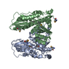
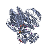
| Structure viewer | Molecule: MolmilJmol/JSmol |
|---|
- Downloads & links
-Download
| PDBx/mmCIF format | 7q6y.cif.gz | 198.3 KB | Display | PDBx/mmCIF format |
|---|---|---|---|---|
| PDB format | pdb7q6y.ent.gz | 157.7 KB | Display | PDB format |
| PDBx/mmJSON format | 7q6y.json.gz | Tree view | PDBx/mmJSON format | |
| Others |  Other downloads Other downloads |
-Validation report
| Arichive directory | https://data.pdbj.org/pub/pdb/validation_reports/q6/7q6yftp://data.pdbj.org/pub/pdb/validation_reports/q6/7q6y | HTTPS FTP |
|---|
-Related structure data
| Related structure data | 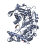 4j0cS S: Starting model for refinement |
|---|---|
| Similar structure data |


-Links
 PDBj
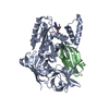
PDBj- Assembly
| Deposited unit | 
| ||||||||||
|---|---|---|---|---|---|---|---|---|---|---|---|
| 1 | 
| ||||||||||
| 2 | 
| ||||||||||
| Unit cell |
|
-Components
| #1: Protein | Mass: 54966.938 Da / Num. of mol.: 2 Source method: isolated from a genetically manipulated source Source: (gene. exp.) Clostridium butyricum (bacteria) / Gene: CBLFYP62_01769, FF104_18625 / Production host: #2: Chemical | ChemComp-PEG /   Mass: 106.120 Da / Num. of mol.: 5 / Source method: isolated from a natural source / Formula: C4H10O3 Mass: 106.120 Da / Num. of mol.: 5 / Source method: isolated from a natural source / Formula: C4H10O3#3: Chemical | ChemComp-EDO /   Mass: 62.068 Da / Num. of mol.: 11 / Source method: isolated from a natural source / Formula: C2H6O2 Mass: 62.068 Da / Num. of mol.: 11 / Source method: isolated from a natural source / Formula: C2H6O2#4: Water | ChemComp-HOH / |  Mass: 18.015 Da / Num. of mol.: 228 / Source method: isolated from a natural source / Formula: H2O Mass: 18.015 Da / Num. of mol.: 228 / Source method: isolated from a natural source / Formula: H2OHas ligand of interest | N | Has protein modification | Y | |
|---|
-Experimental details
-Experiment
| Experiment | Method: X-RAY DIFFRACTION / Number of used crystals: 1 |
|---|
- Sample preparation
| Crystal | Density Matthews: 2.58 Å3/Da / Density % sol: 52.28 % |
|---|---|
| Crystal grow | Temperature: 298.15 K / Method: vapor diffusion, sitting drop / pH: 5 Details: 0.2 M calcium chloride dihydrate 0.1 M sodium acetate, pH 5.0 20 % PEG 6,000 25 mg/mL protein PH range: 4.75-5.75 / Temp details: 25 C |
-Data collection
| Diffraction | Mean temperature: 100 K / Serial crystal experiment: N |
|---|---|
| Diffraction source | Source: SYNCHROTRON / Site: ESRF  / Beamline: ID23-2 / Wavelength: 0.8731 Å / Beamline: ID23-2 / Wavelength: 0.8731 Å |
| Detector | Type: DECTRIS PILATUS3 X 2M / Detector: PIXEL / Date: Sep 18, 2021 |
| Radiation | Protocol: SINGLE WAVELENGTH / Monochromatic (M) / Laue (L): M / Scattering type: x-ray |
| Radiation wavelength | Wavelength: 0.8731 Å / Relative weight: 1 |
| Reflection | Resolution: 2.22→46.45 Å / Num. obs: 56815 / % possible obs: 99.9 % / Redundancy: 13.2 % / Biso Wilson estimate: 43.04 Å2 / Rmerge(I) obs: 0.196 / Net I/σ(I): 11.93 |
| Reflection shell | Resolution: 2.22→2.3 Å / Redundancy: 13.5 % / Rmerge(I) obs: 2.463 / Mean I/σ(I) obs: 1.1 / Num. unique obs: 5560 / % possible all: 99.7 |
- Processing
| Software |
| |||||||||||||||||||||||||||||||||||||||||||||||||||||||||||||||||||||||||||||||||||||||||||||||||||||||||
|---|---|---|---|---|---|---|---|---|---|---|---|---|---|---|---|---|---|---|---|---|---|---|---|---|---|---|---|---|---|---|---|---|---|---|---|---|---|---|---|---|---|---|---|---|---|---|---|---|---|---|---|---|---|---|---|---|---|---|---|---|---|---|---|---|---|---|---|---|---|---|---|---|---|---|---|---|---|---|---|---|---|---|---|---|---|---|---|---|---|---|---|---|---|---|---|---|---|---|---|---|---|---|---|---|---|---|
| Refinement | Method to determine structure: MOLECULAR REPLACEMENT Starting model: 4J0C Resolution: 2.22→46.45 Å / SU ML: 0.334 / Cross valid method: FREE R-VALUE / σ(F): 1.34 / Phase error: 29.825 Stereochemistry target values: GEOSTD + MONOMER LIBRARY + CDL V1.2
| |||||||||||||||||||||||||||||||||||||||||||||||||||||||||||||||||||||||||||||||||||||||||||||||||||||||||
| Solvent computation | Shrinkage radii: 0.9 Å / VDW probe radii: 1.11 Å / Solvent model: FLAT BULK SOLVENT MODEL | |||||||||||||||||||||||||||||||||||||||||||||||||||||||||||||||||||||||||||||||||||||||||||||||||||||||||
| Displacement parameters | Biso mean: 54.34 Å2 | |||||||||||||||||||||||||||||||||||||||||||||||||||||||||||||||||||||||||||||||||||||||||||||||||||||||||
| Refinement step | Cycle: LAST / Resolution: 2.22→46.45 Å
| |||||||||||||||||||||||||||||||||||||||||||||||||||||||||||||||||||||||||||||||||||||||||||||||||||||||||
| Refine LS restraints |
| |||||||||||||||||||||||||||||||||||||||||||||||||||||||||||||||||||||||||||||||||||||||||||||||||||||||||
| LS refinement shell |
|
