 Movie
Movie Controller


+ Open data

- Basic information
| Entry | Database: PDB / ID: 7q5c | ||||||
|---|---|---|---|---|---|---|---|
| Title | Crystal structure of OmpG in space group 96 | ||||||
 Components Components | Outer membrane porin G | ||||||
 Keywords Keywords | MEMBRANE PROTEIN / OmpG / Channel / Membrane / Oligosaccharide | ||||||
| Function / homology |  Function and homology information Function and homology informationcarbohydrate transmembrane transport / oligosaccharide transporting porin activity / maltose transporting porin activity / porin activity / pore complex / monoatomic ion transport / cell outer membrane Similarity search - Function | ||||||
| Biological species |  | ||||||
| Method |  X-RAY DIFFRACTION / SYNCHROTRON / MOLECULAR REPLACEMENT / Resolution: 2.717 Å X-RAY DIFFRACTION / SYNCHROTRON / MOLECULAR REPLACEMENT / Resolution: 2.717 Å | ||||||
 Authors Authors | Nguyen, T.T.M. / Khan, A.R. / Barringer, R. / McManus, J.J. / Race, P.R. | ||||||
| Funding support | European Union, 1items
| ||||||
 Citation Citation | Journal: To Be Published Title: Experimental phase diagrams to optimise OmpG Authors: Nguyen, T.T.M. / Khan, A.R. / Barringer, R. / McManus, J.J. / Race, P.R. | ||||||
| History |
|
- Structure visualization
| Structure viewer | Molecule: MolmilJmol/JSmol |
|---|
- Downloads & links
-Download
| PDBx/mmCIF format | 7q5c.cif.gz | 110.8 KB | Display | PDBx/mmCIF format |
|---|---|---|---|---|
| PDB format | pdb7q5c.ent.gz | Display | PDB format | |
| PDBx/mmJSON format | 7q5c.json.gz | Tree view | PDBx/mmJSON format | |
| Others |  Other downloads Other downloads |
-Validation report
| Summary document | 7q5c_validation.pdf.gz | 799.5 KB | Display | wwPDB validaton report |
|---|---|---|---|---|
| Full document | 7q5c_full_validation.pdf.gz | 806.8 KB | Display | |
| Data in XML | 7q5c_validation.xml.gz | 12.7 KB | Display | |
| Data in CIF | 7q5c_validation.cif.gz | 16.3 KB | Display | |
| Arichive directory | https://data.pdbj.org/pub/pdb/validation_reports/q5/7q5cftp://data.pdbj.org/pub/pdb/validation_reports/q5/7q5c | HTTPS FTP |
-Related structure data
| Related structure data |  2iwvS S: Starting model for refinement |
|---|---|
| Similar structure data |
-Links
 PDBj
PDBj- Assembly
| Deposited unit | 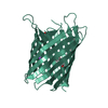
| ||||||||
|---|---|---|---|---|---|---|---|---|---|
| 1 |
| ||||||||
| Unit cell |
| ||||||||
| Components on special symmetry positions |
|
-Components
| #1: Protein | Mass: 32936.527 Da / Num. of mol.: 1 Source method: isolated from a genetically manipulated source Details: Residues showing unresolvable side chains (i.e. with low confidence regarding fit to electron density) have been represented as Alanines in lieu of the bona fide residues. Source: (gene. exp.) Strain: K12 / Gene: ompG, b1319, JW1312 / Production host: | ||||||||
|---|---|---|---|---|---|---|---|---|---|
| #2: Chemical |   Mass: 194.226 Da / Num. of mol.: 2 / Source method: obtained synthetically / Formula: C8H18O5 / Comment: precipitant*YM Mass: 194.226 Da / Num. of mol.: 2 / Source method: obtained synthetically / Formula: C8H18O5 / Comment: precipitant*YM#3: Sugar |   Type: D-saccharide / Mass: 292.369 Da / Num. of mol.: 2 / Source method: obtained synthetically / Formula: C14H28O6 / Feature type: SUBJECT OF INVESTIGATION / Comment: detergent*YM Type: D-saccharide / Mass: 292.369 Da / Num. of mol.: 2 / Source method: obtained synthetically / Formula: C14H28O6 / Feature type: SUBJECT OF INVESTIGATION / Comment: detergent*YM#4: Chemical | ChemComp-NA / |   Mass: 22.990 Da / Num. of mol.: 1 / Source method: isolated from a natural source / Formula: Na Mass: 22.990 Da / Num. of mol.: 1 / Source method: isolated from a natural source / Formula: Na#5: Water | ChemComp-HOH / |  Mass: 18.015 Da / Num. of mol.: 17 / Source method: isolated from a natural source / Formula: H2O Mass: 18.015 Da / Num. of mol.: 17 / Source method: isolated from a natural source / Formula: H2OHas ligand of interest | Y | |
-Experimental details
-Experiment
| Experiment | Method: X-RAY DIFFRACTION / Number of used crystals: 1 |
|---|
- Sample preparation
| Crystal | Density Matthews: 4.41 Å3/Da / Density % sol: 72.08 % |
|---|---|
| Crystal grow | Temperature: 294 K / Method: vapor diffusion / pH: 5.6 Details: 30-60 mM Sodium Citrate, 50-70mM NaCl, 12-14 % (w/v) PEG-3350, 60-65 mM n-octyl-beta-d-glucoside, 6-12 mg/mL protein Temp details: 20/21 celsius |
-Data collection
| Diffraction | Mean temperature: 100 K / Serial crystal experiment: N |
|---|---|
| Diffraction source | Source: SYNCHROTRON / Site: SOLEIL  / Beamline: PROXIMA 2 / Wavelength: 0.97918 Å / Beamline: PROXIMA 2 / Wavelength: 0.97918 Å |
| Detector | Type: DECTRIS EIGER X 9M / Detector: PIXEL / Date: Mar 31, 2019 |
| Radiation | Protocol: SINGLE WAVELENGTH / Monochromatic (M) / Laue (L): M / Scattering type: x-ray |
| Radiation wavelength | Wavelength: 0.97918 Å / Relative weight: 1 |
| Reflection | Resolution: 2.717→67.304 Å / Num. obs: 14670 / % possible obs: 99.7 % / Redundancy: 2 % / Rrim(I) all: 0.0247 / Net I/σ(I): 16.9 |
| Reflection shell | Resolution: 2.717→2.788 Å / Num. unique obs: 980 / Rrim(I) all: 0.83 |
- Processing
| Software |
| |||||||||||||||||||||||||||||||||||||||||||||||||||||||||||||||||||||||||||||||||||||||||||||||||||||||||||||||||||||||||||||||||||||||||||||||||||||||||||
|---|---|---|---|---|---|---|---|---|---|---|---|---|---|---|---|---|---|---|---|---|---|---|---|---|---|---|---|---|---|---|---|---|---|---|---|---|---|---|---|---|---|---|---|---|---|---|---|---|---|---|---|---|---|---|---|---|---|---|---|---|---|---|---|---|---|---|---|---|---|---|---|---|---|---|---|---|---|---|---|---|---|---|---|---|---|---|---|---|---|---|---|---|---|---|---|---|---|---|---|---|---|---|---|---|---|---|---|---|---|---|---|---|---|---|---|---|---|---|---|---|---|---|---|---|---|---|---|---|---|---|---|---|---|---|---|---|---|---|---|---|---|---|---|---|---|---|---|---|---|---|---|---|---|---|---|---|
| Refinement | Method to determine structure: MOLECULAR REPLACEMENT Starting model: 2iwv Resolution: 2.717→67.304 Å / Cor.coef. Fo:Fc: 0.829 / Cor.coef. Fo:Fc free: 0.884 / Cross valid method: FREE R-VALUE / ESU R: 0.518 / ESU R Free: 0.37 Details: Hydrogens have been used in their riding positions for refinement.
| |||||||||||||||||||||||||||||||||||||||||||||||||||||||||||||||||||||||||||||||||||||||||||||||||||||||||||||||||||||||||||||||||||||||||||||||||||||||||||
| Solvent computation | Ion probe radii: 0.8 Å / Shrinkage radii: 0.8 Å / VDW probe radii: 1.2 Å / Solvent model: MASK BULK SOLVENT | |||||||||||||||||||||||||||||||||||||||||||||||||||||||||||||||||||||||||||||||||||||||||||||||||||||||||||||||||||||||||||||||||||||||||||||||||||||||||||
| Displacement parameters | Biso mean: 77.184 Å2
| |||||||||||||||||||||||||||||||||||||||||||||||||||||||||||||||||||||||||||||||||||||||||||||||||||||||||||||||||||||||||||||||||||||||||||||||||||||||||||
| Refinement step | Cycle: LAST / Resolution: 2.717→67.304 Å
| |||||||||||||||||||||||||||||||||||||||||||||||||||||||||||||||||||||||||||||||||||||||||||||||||||||||||||||||||||||||||||||||||||||||||||||||||||||||||||
| Refine LS restraints |
| |||||||||||||||||||||||||||||||||||||||||||||||||||||||||||||||||||||||||||||||||||||||||||||||||||||||||||||||||||||||||||||||||||||||||||||||||||||||||||
| LS refinement shell |
|
