[English] 日本語
Yorodumi
- PDB-7q52: Crystal structure of S/T protein kinase PknG from Mycobacterium t... -

+
Open data


ID or keywords:

Loading...

-
Basic information

Entry
Database: PDB / ID: 7q52
TitleCrystal structure of S/T protein kinase PknG from Mycobacterium tuberculosis in complex with inhibitor L2W
ComponentsSerine/threonine-protein kinase PknG
KeywordsTRANSFERASE / Tuberculosis / inhibitor / L2W
Function / homology
Function and homology information


symbiont-mediated perturbation of host signal transduction pathway / symbiont-mediated perturbation of host defenses / glutamine catabolic process / symbiont-mediated suppression of host innate immune response / glutamate catabolic process / evasion of host immune response / protein autophosphorylation / non-specific serine/threonine protein kinase / response to antibiotic / protein serine kinase activity ...symbiont-mediated perturbation of host signal transduction pathway / symbiont-mediated perturbation of host defenses / glutamine catabolic process / symbiont-mediated suppression of host innate immune response / glutamate catabolic process / evasion of host immune response / protein autophosphorylation / non-specific serine/threonine protein kinase / response to antibiotic / protein serine kinase activity / protein serine/threonine kinase activity / ATP binding / identical protein binding / plasma membrane / cytosol
Similarity search - Function
Protein kinase G, rubredoxin domain / Protein kinase G rubredoxin domain / Protein kinase G, tetratricopeptide repeat containing domain / Protein kinase G tetratricopeptide repeat / Tetratricopeptide-like helical domain superfamily / Serine/threonine-protein kinase, active site / Serine/Threonine protein kinases active-site signature. / Protein kinase domain / Serine/Threonine protein kinases, catalytic domain / Protein kinase domain profile. ...Protein kinase G, rubredoxin domain / Protein kinase G rubredoxin domain / Protein kinase G, tetratricopeptide repeat containing domain / Protein kinase G tetratricopeptide repeat / Tetratricopeptide-like helical domain superfamily / Serine/threonine-protein kinase, active site / Serine/Threonine protein kinases active-site signature. / Protein kinase domain / Serine/Threonine protein kinases, catalytic domain / Protein kinase domain profile. / Protein kinase domain / Protein kinase-like domain superfamily
Similarity search - Domain/homology
Chem-8ZC / : / Serine/threonine-protein kinase PknG
Similarity search - Component
Biological speciesMycobacterium tuberculosis (bacteria)
MethodX-RAY DIFFRACTION / SYNCHROTRON / MOLECULAR REPLACEMENT / Resolution: 2.35 Å
AuthorsDefelipe, L.A. / Burastero, O. / Bento, I. / Garcia-Alai, M.M.
Funding supportEuropean Union, 2items
OrganizationGrant numberCountry
Agencia Nacional de Promocion Cientifica y Tecnologica (FONCYT)PICT2015-2276European Union
H2020 Marie Curie Actions of the European Commission664726European Union
CitationJournal: J.Med.Chem. / Year: 2022
Title: Cosolvent Sites-Based Discovery of Mycobacterium Tuberculosis Protein Kinase G Inhibitors.
Authors: Burastero, O. / Defelipe, L.A. / Gola, G. / Tateosian, N.L. / Lopez, E.D. / Martinena, C.B. / Arcon, J.P. / Traian, M.D. / Wetzler, D.E. / Bento, I. / Barril, X. / Ramirez, J. / Marti, M.A. ...Authors: Burastero, O. / Defelipe, L.A. / Gola, G. / Tateosian, N.L. / Lopez, E.D. / Martinena, C.B. / Arcon, J.P. / Traian, M.D. / Wetzler, D.E. / Bento, I. / Barril, X. / Ramirez, J. / Marti, M.A. / Garcia-Alai, M.M. / Turjanski, A.G.
History
DepositionNov 2, 2021Deposition site: PDBE / Processing site: PDBE
Revision 1.0Jun 22, 2022Provider: repository / Type: Initial release
Revision 1.1Jul 6, 2022Group: Database references / Category: citation / citation_author
Item: _citation.country / _citation.journal_abbrev ..._citation.country / _citation.journal_abbrev / _citation.journal_id_ASTM / _citation.journal_id_CSD / _citation.journal_id_ISSN / _citation.pdbx_database_id_PubMed / _citation.title / _citation.year / _citation_author.identifier_ORCID / _citation_author.name
Revision 1.2Aug 10, 2022Group: Database references / Category: citation / citation_author
Item: _citation.journal_volume / _citation.page_first ..._citation.journal_volume / _citation.page_first / _citation.page_last / _citation_author.identifier_ORCID
Revision 1.3Jan 31, 2024Group: Data collection / Refinement description
Category: chem_comp_atom / chem_comp_bond / pdbx_initial_refinement_model

-
Structure visualization

Structure viewerMolecule:
MolmilJmol/JSmol

Downloads & links

-
Assembly

Deposited unit
AAA: Serine/threonine-protein kinase PknG
hetero molecules


Theoretical massNumber of molelcules
Total (without water)36,7345
Polymers36,3351
Non-polymers3994
Water52229
1


  • Idetical with deposited unit
  • defined by author&software
  • Evidence: gel filtration
TypeNameSymmetry operationNumber
identity operation1_555x,y,z1
Buried area250 Å2
ΔGint-19 kcal/mol
Surface area15370 Å2
MethodPISA
Unit cell
Length a, b, c (Å)77.327, 37.016, 106.781
Angle α, β, γ (deg.)90.000, 105.500, 90.000
Int Tables number5
Space group name H-MC121

-
Components

#1: Protein Serine/threonine-protein kinase PknG


Mass: 36335.277 Da / Num. of mol.: 1
Source method: isolated from a genetically manipulated source
Source: (gene. exp.) Mycobacterium tuberculosis (strain ATCC 25618 / H37Rv) (bacteria)
Strain: ATCC 25618 / H37Rv / Gene: pknG, Rv0410c, MTCY22G10.06c / Production host: Escherichia coli BL21(DE3) (bacteria)
References: UniProt: P9WI73, non-specific serine/threonine protein kinase
#2: Chemical ChemComp-FE / FE (III) ION


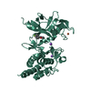
Mass: 55.845 Da / Num. of mol.: 1 / Source method: obtained synthetically / Formula: Fe
#3: Chemical ChemComp-8ZC / 2-azanyl-3-(4-fluorophenyl)carbonyl-indolizine-1-carboxamide


Mass: 297.284 Da / Num. of mol.: 1 / Source method: obtained synthetically / Formula: C16H12FN3O2 / Feature type: SUBJECT OF INVESTIGATION
#4: Chemical ChemComp-NA / SODIUM ION


Mass: 22.990 Da / Num. of mol.: 2 / Source method: obtained synthetically / Formula: Na
#5: Water ChemComp-HOH / water


Mass: 18.015 Da / Num. of mol.: 29 / Source method: isolated from a natural source / Formula: H2O
Has ligand of interestY

-
Experimental details

-
Experiment

ExperimentMethod: X-RAY DIFFRACTION / Number of used crystals: 1

-
Sample preparation

CrystalDensity Matthews: 2.03 Å3/Da / Density % sol: 39.3 %
Crystal growTemperature: 292 K / Method: vapor diffusion, sitting drop / pH: 8.5
Details: Mother Liquor: 0.2M CaCl2, 0.2 M TRIS, pH 8.5 and 20 % (w/v) PEG 4000

-
Data collection

DiffractionMean temperature: 196 K / Serial crystal experiment: N
Diffraction sourceSource: SYNCHROTRON / Site: PETRA III, EMBL c/o DESY / Beamline: P14 (MX2) / Wavelength: 0.9763 Å
DetectorType: DECTRIS EIGER2 X 16M / Detector: PIXEL / Date: Oct 4, 2021
RadiationMonochromator: Si(111) / Protocol: SINGLE WAVELENGTH / Monochromatic (M) / Laue (L): M / Scattering type: x-ray
Radiation wavelengthWavelength: 0.9763 Å / Relative weight: 1
ReflectionResolution: 2.35→51.5 Å / Num. obs: 12303 / % possible obs: 98.4 % / Redundancy: 6.8 % / Biso Wilson estimate: 62.8 Å2 / CC1/2: 0.999 / Net I/σ(I): 1.67
Reflection shellResolution: 2.35→2.43 Å / Redundancy: 6.9 % / Num. unique obs: 1180 / CC1/2: 0.699 / % possible all: 98.1

-
Processing

Software
NameVersionClassification
REFMAC5.8.0267refinement
XDSVERSION Feb 5, 2021data reduction
Aimless0.7.7data scaling
MOLREP11.7.03phasing
RefinementMethod to determine structure: MOLECULAR REPLACEMENT
Starting model: 4Y12
Resolution: 2.35→51.502 Å / Cor.coef. Fo:Fc: 0.964 / Cor.coef. Fo:Fc free: 0.938 / WRfactor Rfree: 0.276 / WRfactor Rwork: 0.218 / SU B: 16.055 / SU ML: 0.345 / Average fsc free: 0.7643 / Average fsc work: 0.8027 / Cross valid method: FREE R-VALUE / ESU R: 0.611 / ESU R Free: 0.296 / Details: Hydrogens have not been used
RfactorNum. reflection% reflection
Rfree0.2676 627 5.12 %
Rwork0.215 11619 -
all0.218 --
obs-12246 98.377 %
Solvent computationIon probe radii: 0.8 Å / Shrinkage radii: 0.8 Å / VDW probe radii: 1.2 Å / Solvent model: MASK BULK SOLVENT
Displacement parametersBiso mean: 74.413 Å2
Baniso -1Baniso -2Baniso -3
1-3.035 Å20 Å21.561 Å2
2--1.17 Å20 Å2
3----4.395 Å2
Refinement stepCycle: LAST / Resolution: 2.35→51.502 Å
ProteinNucleic acidLigandSolventTotal
Num. atoms2401 0 25 29 2455
Refine LS restraints
Refine-IDTypeDev idealDev ideal targetNumber
X-RAY DIFFRACTIONr_bond_refined_d0.0040.0122490
X-RAY DIFFRACTIONr_angle_refined_deg1.2461.643395
X-RAY DIFFRACTIONr_dihedral_angle_1_deg7.15312
X-RAY DIFFRACTIONr_dihedral_angle_2_deg30.72121.811127
X-RAY DIFFRACTIONr_dihedral_angle_3_deg15.89315390
X-RAY DIFFRACTIONr_dihedral_angle_4_deg10.9751518
X-RAY DIFFRACTIONr_chiral_restr0.0920.2314
X-RAY DIFFRACTIONr_gen_planes_refined0.0050.021932
X-RAY DIFFRACTIONr_nbd_refined0.2270.21112
X-RAY DIFFRACTIONr_nbtor_refined0.3120.21713
X-RAY DIFFRACTIONr_xyhbond_nbd_refined0.1410.292
X-RAY DIFFRACTIONr_metal_ion_refined0.1620.21
X-RAY DIFFRACTIONr_symmetry_nbd_refined0.280.264
X-RAY DIFFRACTIONr_symmetry_xyhbond_nbd_refined0.3110.25
X-RAY DIFFRACTIONr_mcbond_it5.1617.3021251
X-RAY DIFFRACTIONr_mcangle_it7.62610.9391562
X-RAY DIFFRACTIONr_scbond_it5.8277.6051239
X-RAY DIFFRACTIONr_scangle_it8.40111.2281833
X-RAY DIFFRACTIONr_lrange_it12.976136.89410396
LS refinement shell
Resolution (Å)Rfactor RfreeNum. reflection RfreeRfactor RworkNum. reflection RworkRefine-ID% reflection obs (%)
2.35-2.4110.325420.374848X-RAY DIFFRACTION98.4513
2.411-2.4770.424460.367824X-RAY DIFFRACTION98.5277
2.477-2.5490.395460.354806X-RAY DIFFRACTION98.6111
2.549-2.6270.544360.362777X-RAY DIFFRACTION98.5455
2.627-2.7130.426550.319765X-RAY DIFFRACTION98.7952
2.713-2.8090.314350.321743X-RAY DIFFRACTION98.8564
2.809-2.9140.43450.306692X-RAY DIFFRACTION99.5946
2.914-3.0330.343400.28696X-RAY DIFFRACTION98.5274
3.033-3.1680.469250.246657X-RAY DIFFRACTION97.9885
3.168-3.3230.314330.248643X-RAY DIFFRACTION98.5423
3.323-3.5020.306350.268582X-RAY DIFFRACTION97.7813
3.502-3.7140.235280.241567X-RAY DIFFRACTION95.9677
3.714-3.970.264350.205536X-RAY DIFFRACTION98.11
3.97-4.2880.289260.178499X-RAY DIFFRACTION98.3146
4.288-4.6960.206210.144459X-RAY DIFFRACTION98.3607
4.696-5.2490.156150.133431X-RAY DIFFRACTION98.022
5.249-6.0590.161180.178362X-RAY DIFFRACTION97.6864
6.059-7.4140.233190.187330X-RAY DIFFRACTION99.1477
7.414-10.4580.195210.134250X-RAY DIFFRACTION99.2674

+
About Yorodumi

-
News

-
Feb 9, 2022. New format data for meta-information of EMDB entries

New format data for meta-information of EMDB entries

  • Version 3 of the EMDB header file is now the official format.
  • The previous official version 1.9 will be removed from the archive.

Related info.:EMDB header

External links:wwPDB to switch to version 3 of the EMDB data model

-
Aug 12, 2020. Covid-19 info

Covid-19 info

URL: https://pdbjlvh1.pdbj.org/emnavi/covid19.php

New page: Covid-19 featured information page in EM Navigator.

Related info.:Covid-19 info / Mar 5, 2020. Novel coronavirus structure data

+
Mar 5, 2020. Novel coronavirus structure data

Novel coronavirus structure data

Related info.:Yorodumi Speices / Aug 12, 2020. Covid-19 info

External links:COVID-19 featured content - PDBj / Molecule of the Month (242):Coronavirus Proteases

+
Jan 31, 2019. EMDB accession codes are about to change! (news from PDBe EMDB page)

EMDB accession codes are about to change! (news from PDBe EMDB page)

  • The allocation of 4 digits for EMDB accession codes will soon come to an end. Whilst these codes will remain in use, new EMDB accession codes will include an additional digit and will expand incrementally as the available range of codes is exhausted. The current 4-digit format prefixed with “EMD-” (i.e. EMD-XXXX) will advance to a 5-digit format (i.e. EMD-XXXXX), and so on. It is currently estimated that the 4-digit codes will be depleted around Spring 2019, at which point the 5-digit format will come into force.
  • The EM Navigator/Yorodumi systems omit the EMD- prefix.

Related info.:Q: What is EMD? / ID/Accession-code notation in Yorodumi/EM Navigator

External links:EMDB Accession Codes are Changing Soon! / Contact to PDBj

+
Jul 12, 2017. Major update of PDB

Major update of PDB

  • wwPDB released updated PDB data conforming to the new PDBx/mmCIF dictionary.
  • This is a major update changing the version number from 4 to 5, and with Remediation, in which all the entries are updated.
  • In this update, many items about electron microscopy experimental information are reorganized (e.g. em_software).
  • Now, EM Navigator and Yorodumi are based on the updated data.

External links:wwPDB Remediation / Enriched Model Files Conforming to OneDep Data Standards Now Available in the PDB FTP Archive

-
Yorodumi

Thousand views of thousand structures

  • Yorodumi is a browser for structure data from EMDB, PDB, SASBDB, etc.
  • This page is also the successor to EM Navigator detail page, and also detail information page/front-end page for Omokage search.
  • The word "yorodu" (or yorozu) is an old Japanese word meaning "ten thousand". "mi" (miru) is to see.

Related info.:EMDB / PDB / SASBDB / Comparison of 3 databanks / Yorodumi Search / Aug 31, 2016. New EM Navigator & Yorodumi / Yorodumi Papers / Jmol/JSmol / Function and homology information / Changes in new EM Navigator and Yorodumi

Read more