 ムービー
ムービー コントローラー


+ データを開く

- 基本情報
基本情報
| 登録情報 | データベース: PDB / ID: 7q3v | ||||||||||||
|---|---|---|---|---|---|---|---|---|---|---|---|---|---|
| タイトル | Re-refined structure of a type III antifreeze protein isoform HPLC 12 | ||||||||||||
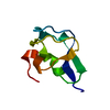
 要素 要素 | Type-3 ice-structuring protein HPLC 12 | ||||||||||||
 キーワード キーワード | ANTIFREEZE PROTEIN / restrained MD refinement scheme / Amber / crystal unit cell model / ensemble refinement | ||||||||||||
| 機能・相同性 | Antifreeze, type III / Antifreeze-like/N-acetylneuraminic acid synthase C-terminal / Antifreeze protein-like domain profile. / Antifreeze-like/N-acetylneuraminic acid synthase C-terminal domain superfamily / extracellular region / Type-3 ice-structuring protein HPLC 12 機能・相同性情報 機能・相同性情報 | ||||||||||||
| 生物種 |  Zoarces americanus (魚類) Zoarces americanus (魚類) | ||||||||||||
| 手法 |  X線回折 / 分子置換 / 解像度: 1.9 Å X線回折 / 分子置換 / 解像度: 1.9 Å | ||||||||||||
 データ登録者 データ登録者 | Mikhailovskii, O. / Xue, Y. / Jia, Z. / Skrynnikov, N.R. | ||||||||||||
| 資金援助 |  ロシア, ロシア,  中国, 3件 中国, 3件
| ||||||||||||
 引用 引用 | ジャーナル: Iucrj / 年: 2022 タイトル: Modeling a unit cell: crystallographic refinement procedure using the biomolecular MD simulation platform Amber. 著者: Mikhailovskii, O. / Xue, Y. / Skrynnikov, N.R. | ||||||||||||
| 履歴 |
|
- 構造の表示
構造の表示
| 構造ビューア | 分子: MolmilJmol/JSmol |
|---|
- ダウンロードとリンク
-ダウンロード
| PDBx/mmCIF形式 | 7q3v.cif.gz | 106.4 KB | 表示 | PDBx/mmCIF形式 |
|---|---|---|---|---|
| PDB形式 | pdb7q3v.ent.gz | 83.8 KB | 表示 | PDB形式 |
| PDBx/mmJSON形式 | 7q3v.json.gz | ツリー表示 | PDBx/mmJSON形式 | |
| その他 |  その他のダウンロード その他のダウンロード |
-検証レポート
| 文書・要旨 | 7q3v_validation.pdf.gz | 440 KB | 表示 | wwPDB検証レポート |
|---|---|---|---|---|
| 文書・詳細版 | 7q3v_full_validation.pdf.gz | 440.9 KB | 表示 | |
| XML形式データ | 7q3v_validation.xml.gz | 12.3 KB | 表示 | |
| CIF形式データ | 7q3v_validation.cif.gz | 18 KB | 表示 | |
| アーカイブディレクトリ | https://data.pdbj.org/pub/pdb/validation_reports/q3/7q3vftp://data.pdbj.org/pub/pdb/validation_reports/q3/7q3v | HTTPS FTP |
-関連構造データ
| 関連構造データ | 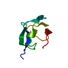 1msiS S: 精密化の開始モデル |
|---|---|
| 類似構造データ |
-リンク
 PDBj
PDBj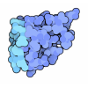
- 集合体
集合体
| 登録構造単位 | 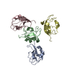
| ||||||||
|---|---|---|---|---|---|---|---|---|---|
| 1 | 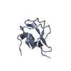
| ||||||||
| 2 | 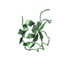
| ||||||||
| 3 | 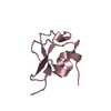
| ||||||||
| 4 | 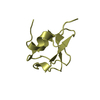
| ||||||||
| 単位格子 |
|
-要素
| #1: タンパク質 | 分子量: 7056.444 Da / 分子数: 4 / 由来タイプ: 組換発現 / 由来: (組換発現) Zoarces americanus (魚類) / 発現宿主:  #2: 水 | ChemComp-HOH / |  分子量: 18.015 Da / 分子数: 155 / 由来タイプ: 天然 / 式: H2O 分子量: 18.015 Da / 分子数: 155 / 由来タイプ: 天然 / 式: H2O |
|---|
-実験情報
-実験
| 実験 | 手法: X線回折 / 使用した結晶の数: 1 |
|---|
- 試料調製
試料調製
| 結晶 | マシュー密度: 2.09 Å3/Da / 溶媒含有率: 30 % |
|---|---|
| 結晶化 | 温度: 293 K / 手法: 蒸気拡散法, ハンギングドロップ法 詳細: 50-55% AMMONIUM SULFATE, 0.1 M SODIUM ACETATE PH 4-4.5 |
-データ収集
| 回折 | 平均測定温度: 295 K / Serial crystal experiment: N |
|---|---|
| 放射光源 | 由来: 回転陽極 / タイプ: RIGAKU / 波長: 1.5418 Å |
| 検出器 | タイプ: MARRESEARCH / 検出器: IMAGE PLATE / 日付: 1996年7月11日 |
| 放射 | プロトコル: SINGLE WAVELENGTH / 単色(M)・ラウエ(L): M / 散乱光タイプ: x-ray |
| 放射波長 | 波長: 1.5418 Å / 相対比: 1 |
| 反射 | 解像度: 1.9→14.86 Å / Num. obs: 19230 / % possible obs: 99.1 % / 冗長度: 3.8 % / Rsym value: 0.077 / Net I/σ(I): 7.8 |
| 反射 シェル | 解像度: 1.9→1.97 Å / Rsym value: 0.306 / % possible all: 98.3 |
- 解析
解析
| ソフトウェア |
| ||||||||||||||||||||
|---|---|---|---|---|---|---|---|---|---|---|---|---|---|---|---|---|---|---|---|---|---|
| 精密化 | 構造決定の手法: 分子置換 開始モデル: 1MSI 解像度: 1.9→14.86 Å / 交差検証法: FREE R-VALUE / 位相誤差: 21.18 詳細: The original structure, 2MSI, has been expanded into a crystal unit cell (containing four protein chains); the resulting model was subsequently refined through a specially designed ...詳細: The original structure, 2MSI, has been expanded into a crystal unit cell (containing four protein chains); the resulting model was subsequently refined through a specially designed constrained MD run using a customized version of the program Amber. Note: ordered water molecules are added after the refinement using the standard water-picking facility of phenix.refine.
| ||||||||||||||||||||
| 溶媒の処理 | 減衰半径: 0.9 Å / VDWプローブ半径: 1.11 Å | ||||||||||||||||||||
| 原子変位パラメータ | Biso max: 61.39 Å2 / Biso mean: 20.1134 Å2 / Biso min: 6.09 Å2 | ||||||||||||||||||||
| 精密化ステップ | サイクル: LAST / 解像度: 1.9→14.86 Å
| ||||||||||||||||||||
| LS精密化 シェル | 解像度: 1.9→1.96 Å
|
