 Movie
Movie Controller

[English] 日本語
 Yorodumi
- PDB-7q0q: Acetyltrasferase(3) type IIIa in complex with 3-N-methyl-nemycin B -
+ Open data

- Basic information
| Entry | Database: PDB / ID: 7q0q | |||||||||
|---|---|---|---|---|---|---|---|---|---|---|
| Title | Acetyltrasferase(3) type IIIa in complex with 3-N-methyl-nemycin B | |||||||||
 Components Components | Aminoglycoside N(3)-acetyltransferase III | |||||||||
 Keywords Keywords | ANTIBIOTIC / acetyltrasferase / aminoglycosides / inhibitor / complex | |||||||||
| Function / homology |  Function and homology information Function and homology information2-deoxystreptamine metabolic process / aminoglycoside 3-N-acetyltransferase / antibiotic metabolic process / aminoglycoside 3-N-acetyltransferase activity / acetyl-CoA catabolic process / coenzyme A binding / cellular response to antibiotic / protein homodimerization activity / identical protein binding Similarity search - Function | |||||||||
| Biological species |   Pseudomonas aeruginosa (bacteria) Pseudomonas aeruginosa (bacteria) | |||||||||
| Method |  X-RAY DIFFRACTION / SYNCHROTRON / MOLECULAR REPLACEMENT / Resolution: 1.96 Å X-RAY DIFFRACTION / SYNCHROTRON / MOLECULAR REPLACEMENT / Resolution: 1.96 Å | |||||||||
 Authors Authors | Pontillo, N. / Guskov, A. | |||||||||
| Funding support | European Union, 2items
| |||||||||
 Citation Citation | Journal: To Be Published Title: The 3-N-alkylation of the neomycin B outmaneuvers the aminoglycoside resistant enzyme acetyltransferase(3)IIIa via an unexpected mechanism Authors: Pontillo, N. / Warszawik, E.M. / Partipilo, M. / Stetsenko, A. / Loznik, M. / Yang, X. / Slotboom, D.J. / Guskov, A. / Herrmann, A. | |||||||||
| History |
|
- Structure visualization
| Structure viewer | Molecule: MolmilJmol/JSmol |
|---|
- Downloads & links
-Download
| PDBx/mmCIF format | 7q0q.cif.gz | 157.9 KB | Display | PDBx/mmCIF format |
|---|---|---|---|---|
| PDB format | pdb7q0q.ent.gz | 98.9 KB | Display | PDB format |
| PDBx/mmJSON format | 7q0q.json.gz | Tree view | PDBx/mmJSON format | |
| Others |  Other downloads Other downloads |
-Validation report
| Arichive directory | https://data.pdbj.org/pub/pdb/validation_reports/q0/7q0qftp://data.pdbj.org/pub/pdb/validation_reports/q0/7q0q | HTTPS FTP |
|---|
-Related structure data
| Related structure data |  7q1xC 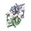 5ht0S  6mb9S  6np4S 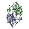 7kesS S: Starting model for refinement C: citing same article ( |
|---|---|
| Similar structure data |
-Links
 PDBj
PDBj- Assembly
| Deposited unit | 
| ||||||||||||
|---|---|---|---|---|---|---|---|---|---|---|---|---|---|
| 1 |
| ||||||||||||
| Unit cell |
|
-Components
-Protein , 1 types, 2 molecules AB
| #1: Protein | Mass: 29429.445 Da / Num. of mol.: 2 Source method: isolated from a genetically manipulated source Source: (gene. exp.) Pseudomonas aeruginosa (bacteria) / Gene: aacC3Production host: References: UniProt: P29808, aminoglycoside 3-N-acetyltransferase |
|---|
-Non-polymers , 6 types, 501 molecules 

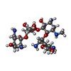


| #2: Chemical |  Mass: 92.094 Da / Num. of mol.: 2 / Source method: obtained synthetically / Formula: C3H8O3 Mass: 92.094 Da / Num. of mol.: 2 / Source method: obtained synthetically / Formula: C3H8O3#3: Chemical | ChemComp-ACT / |  Mass: 59.044 Da / Num. of mol.: 1 / Source method: obtained synthetically / Formula: C2H3O2 Mass: 59.044 Da / Num. of mol.: 1 / Source method: obtained synthetically / Formula: C2H3O2#4: Chemical | ChemComp-8I5 / |  Mass: 628.670 Da / Num. of mol.: 1 / Source method: obtained synthetically / Formula: C24H48N6O13 / Feature type: SUBJECT OF INVESTIGATION Mass: 628.670 Da / Num. of mol.: 1 / Source method: obtained synthetically / Formula: C24H48N6O13 / Feature type: SUBJECT OF INVESTIGATION#5: Chemical |  Mass: 35.453 Da / Num. of mol.: 3 / Source method: obtained synthetically / Formula: Cl Mass: 35.453 Da / Num. of mol.: 3 / Source method: obtained synthetically / Formula: Cl#6: Chemical | ChemComp-PEG / |  Mass: 106.120 Da / Num. of mol.: 1 / Source method: obtained synthetically / Formula: C4H10O3 Mass: 106.120 Da / Num. of mol.: 1 / Source method: obtained synthetically / Formula: C4H10O3#7: Water | ChemComp-HOH / | Mass: 18.015 Da / Num. of mol.: 493 / Source method: isolated from a natural source / Formula: H2O |
|---|
-Details
| Has ligand of interest | Y |
|---|
-Experimental details
-Experiment
| Experiment | Method: X-RAY DIFFRACTION / Number of used crystals: 1 |
|---|
- Sample preparation
| Crystal | Density Matthews: 2.53 Å3/Da / Density % sol: 51.34 % / Description: orthorombic |
|---|---|
| Crystal grow | Temperature: 298 K / Method: vapor diffusion, sitting drop Details: 7-14% PEG 10,000, 0.1 M Na Acetate, 0.1 M Bis-Tris pH 5.5 |
-Data collection
| Diffraction | Mean temperature: 110 K / Serial crystal experiment: N |
|---|---|
| Diffraction source | Source: SYNCHROTRON / Site: PETRA III, EMBL c/o DESY  / Beamline: P13 (MX1) / Wavelength: 0.987 Å / Beamline: P13 (MX1) / Wavelength: 0.987 Å |
| Detector | Type: DECTRIS EIGER R 4M / Detector: PIXEL / Date: Nov 11, 2018 |
| Radiation | Protocol: SINGLE WAVELENGTH / Monochromatic (M) / Laue (L): M / Scattering type: x-ray |
| Radiation wavelength | Wavelength: 0.987 Å / Relative weight: 1 |
| Reflection | Resolution: 1.96→19.67 Å / Num. obs: 43209 / % possible obs: 99.13 % / Redundancy: 7.2 % / Biso Wilson estimate: 30.1 Å2 / CC1/2: 0.996 / Rmerge(I) obs: 0.1743 / Net I/σ(I): 8.04 |
| Reflection shell | Resolution: 1.96→2.03 Å / Num. unique obs: 3999 / CC1/2: 0.551 |
- Processing
| Software |
| ||||||||||||||||||||||||||||||||||||||||||||||||||||||||||||||||||||||||||||||||||||||||||||||||||||||||||||||||||||||||||||||||||||||||||||||||||||||||||||||||||||||||||||||||||||||||||||||||||||||||||||||||||
|---|---|---|---|---|---|---|---|---|---|---|---|---|---|---|---|---|---|---|---|---|---|---|---|---|---|---|---|---|---|---|---|---|---|---|---|---|---|---|---|---|---|---|---|---|---|---|---|---|---|---|---|---|---|---|---|---|---|---|---|---|---|---|---|---|---|---|---|---|---|---|---|---|---|---|---|---|---|---|---|---|---|---|---|---|---|---|---|---|---|---|---|---|---|---|---|---|---|---|---|---|---|---|---|---|---|---|---|---|---|---|---|---|---|---|---|---|---|---|---|---|---|---|---|---|---|---|---|---|---|---|---|---|---|---|---|---|---|---|---|---|---|---|---|---|---|---|---|---|---|---|---|---|---|---|---|---|---|---|---|---|---|---|---|---|---|---|---|---|---|---|---|---|---|---|---|---|---|---|---|---|---|---|---|---|---|---|---|---|---|---|---|---|---|---|---|---|---|---|---|---|---|---|---|---|---|---|---|---|---|---|---|
| Refinement | Method to determine structure: MOLECULAR REPLACEMENT Starting model: 5ht0,7kes,6mb9,6np4 Resolution: 1.96→19.87 Å / SU ML: 0.2853 / Cross valid method: FREE R-VALUE / σ(F): 1.91 / Phase error: 26.2587 Stereochemistry target values: GeoStd + Monomer Library + CDL v1.2
| ||||||||||||||||||||||||||||||||||||||||||||||||||||||||||||||||||||||||||||||||||||||||||||||||||||||||||||||||||||||||||||||||||||||||||||||||||||||||||||||||||||||||||||||||||||||||||||||||||||||||||||||||||
| Solvent computation | Shrinkage radii: 0.9 Å / VDW probe radii: 1.11 Å / Solvent model: FLAT BULK SOLVENT MODEL | ||||||||||||||||||||||||||||||||||||||||||||||||||||||||||||||||||||||||||||||||||||||||||||||||||||||||||||||||||||||||||||||||||||||||||||||||||||||||||||||||||||||||||||||||||||||||||||||||||||||||||||||||||
| Displacement parameters | Biso mean: 34.43 Å2 | ||||||||||||||||||||||||||||||||||||||||||||||||||||||||||||||||||||||||||||||||||||||||||||||||||||||||||||||||||||||||||||||||||||||||||||||||||||||||||||||||||||||||||||||||||||||||||||||||||||||||||||||||||
| Refinement step | Cycle: LAST / Resolution: 1.96→19.87 Å
| ||||||||||||||||||||||||||||||||||||||||||||||||||||||||||||||||||||||||||||||||||||||||||||||||||||||||||||||||||||||||||||||||||||||||||||||||||||||||||||||||||||||||||||||||||||||||||||||||||||||||||||||||||
| Refine LS restraints |
| ||||||||||||||||||||||||||||||||||||||||||||||||||||||||||||||||||||||||||||||||||||||||||||||||||||||||||||||||||||||||||||||||||||||||||||||||||||||||||||||||||||||||||||||||||||||||||||||||||||||||||||||||||
| LS refinement shell |
|
