 Movie
Movie Controller

[English] 日本語
 Yorodumi
- PDB-7pxy: Crystal structure of Arabidopsis thaliana 5-enol-pyruvyl-shikimat... -
+ Open data

- Basic information
| Entry | Database: PDB / ID: 7pxy | ||||||
|---|---|---|---|---|---|---|---|
| Title | Crystal structure of Arabidopsis thaliana 5-enol-pyruvyl-shikimate-3-phosphate synthase (EPSPS) in open conformation | ||||||
 Components Components | 3-phosphoshikimate 1-carboxyvinyltransferase, chloroplastic | ||||||
 Keywords Keywords | PLANT PROTEIN / glyphosate / herbicide design / EPSP synthase | ||||||
| Function / homology |  Function and homology information Function and homology information3-phosphoshikimate 1-carboxyvinyltransferase / 3-phosphoshikimate 1-carboxyvinyltransferase activity / chorismate biosynthetic process / aromatic amino acid biosynthetic process / chloroplast stroma / amino acid biosynthetic process / chloroplast Similarity search - Function | ||||||
| Biological species |  | ||||||
| Method |  X-RAY DIFFRACTION / SYNCHROTRON / MOLECULAR REPLACEMENT / Resolution: 1.4 Å X-RAY DIFFRACTION / SYNCHROTRON / MOLECULAR REPLACEMENT / Resolution: 1.4 Å | ||||||
 Authors Authors | Ruszkowski, M. | ||||||
| Funding support |  United States, 1items United States, 1items
| ||||||
 Citation Citation | Journal: Comput Struct Biotechnol J / Year: 2022 Title: Deciphering the structure of Arabidopsis thaliana 5- enol -pyruvyl-shikimate-3-phosphate synthase: An essential step toward the discovery of novel inhibitors to supersede glyphosate. Authors: Ruszkowski, M. / Forlani, G. | ||||||
| History |
|
- Structure visualization
| Structure viewer | Molecule: MolmilJmol/JSmol |
|---|
- Downloads & links
-Download
| PDBx/mmCIF format | 7pxy.cif.gz | 241.6 KB | Display | PDBx/mmCIF format |
|---|---|---|---|---|
| PDB format | pdb7pxy.ent.gz | 159.9 KB | Display | PDB format |
| PDBx/mmJSON format | 7pxy.json.gz | Tree view | PDBx/mmJSON format | |
| Others |  Other downloads Other downloads |
-Validation report
| Arichive directory | https://data.pdbj.org/pub/pdb/validation_reports/px/7pxyftp://data.pdbj.org/pub/pdb/validation_reports/px/7pxy | HTTPS FTP |
|---|
-Related structure data
| Related structure data |  3nvsS S: Starting model for refinement |
|---|---|
| Similar structure data |
-Links
 PDBj
PDBj
- Assembly
| Deposited unit | 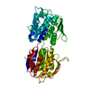
| |||||||||||||||
|---|---|---|---|---|---|---|---|---|---|---|---|---|---|---|---|---|
| 1 |
| |||||||||||||||
| Unit cell |
| |||||||||||||||
| Components on special symmetry positions |
|
-Components
| #1: Protein | Mass: 47872.664 Da / Num. of mol.: 1 Source method: isolated from a genetically manipulated source Source: (gene. exp.)  References: UniProt: P05466, 3-phosphoshikimate 1-carboxyvinyltransferase |
|---|---|
| #2: Chemical | ChemComp-CL /   Mass: 35.453 Da / Num. of mol.: 1 / Source method: obtained synthetically / Formula: Cl Mass: 35.453 Da / Num. of mol.: 1 / Source method: obtained synthetically / Formula: Cl |
| #3: Chemical | ChemComp-MG /   Mass: 24.305 Da / Num. of mol.: 1 / Source method: obtained synthetically / Formula: Mg Mass: 24.305 Da / Num. of mol.: 1 / Source method: obtained synthetically / Formula: Mg |
| #4: Water | ChemComp-HOH /  Mass: 18.015 Da / Num. of mol.: 478 / Source method: isolated from a natural source / Formula: H2O Mass: 18.015 Da / Num. of mol.: 478 / Source method: isolated from a natural source / Formula: H2O |
| Has ligand of interest | N |
-Experimental details
-Experiment
| Experiment | Method: X-RAY DIFFRACTION / Number of used crystals: 1 |
|---|
- Sample preparation
| Crystal | Density Matthews: 2.41 Å3/Da / Density % sol: 48.88 % |
|---|---|
| Crystal grow | Temperature: 292 K / Method: vapor diffusion, sitting drop / pH: 7.5 Details: 75% of Index G12 (0.2 M Magnesium chloride hexahydrate 0.1 M HEPES pH 7.5 25% w/v Polyethylene glycol 3,350) + 10 mM glyphosate 2+2 drop Cryoprotection: Index G12+20% ethylene glycol; 0.5 ul ...Details: 75% of Index G12 (0.2 M Magnesium chloride hexahydrate 0.1 M HEPES pH 7.5 25% w/v Polyethylene glycol 3,350) + 10 mM glyphosate 2+2 drop Cryoprotection: Index G12+20% ethylene glycol; 0.5 ul of 200 mM glyphosate was added to 10 ul of cryo solution |
-Data collection
| Diffraction | Mean temperature: 100 K / Serial crystal experiment: N |
|---|---|
| Diffraction source | Source: SYNCHROTRON / Site: APS / Beamline: 22-ID / Wavelength: 1 Å |
| Detector | Type: DECTRIS EIGER X 16M / Detector: PIXEL / Date: Oct 20, 2018 |
| Radiation | Monochromator: Si(111) / Protocol: SINGLE WAVELENGTH / Monochromatic (M) / Laue (L): M / Scattering type: x-ray |
| Radiation wavelength | Wavelength: 1 Å / Relative weight: 1 |
| Reflection | Resolution: 1.4→80 Å / Num. obs: 92087 / % possible obs: 99.9 % / Redundancy: 18.5 % / Biso Wilson estimate: 22.24 Å2 / CC1/2: 0.999 / Rmerge(I) obs: 0.066 / Net I/σ(I): 21.8 |
| Reflection shell | Resolution: 1.4→1.48 Å / Redundancy: 18.7 % / Rmerge(I) obs: 1.4 / Mean I/σ(I) obs: 2.1 / Num. unique obs: 14710 / CC1/2: 0.784 / % possible all: 99.7 |
- Processing
| Software |
| ||||||||||||||||||||||||||||||||||||||||||||||||||||||||
|---|---|---|---|---|---|---|---|---|---|---|---|---|---|---|---|---|---|---|---|---|---|---|---|---|---|---|---|---|---|---|---|---|---|---|---|---|---|---|---|---|---|---|---|---|---|---|---|---|---|---|---|---|---|---|---|---|---|
| Refinement | Method to determine structure: MOLECULAR REPLACEMENT Starting model: 3nvs Resolution: 1.4→47.08 Å / SU ML: 0.1729 / Cross valid method: FREE R-VALUE / σ(F): 1.35 / Phase error: 21.5411 Stereochemistry target values: GeoStd + Monomer Library + CDL v1.2 Details: Anisotropic refinement of ADPs,
| ||||||||||||||||||||||||||||||||||||||||||||||||||||||||
| Solvent computation | Shrinkage radii: 0.9 Å / VDW probe radii: 1.11 Å / Solvent model: FLAT BULK SOLVENT MODEL | ||||||||||||||||||||||||||||||||||||||||||||||||||||||||
| Displacement parameters | Biso mean: 31.48 Å2 | ||||||||||||||||||||||||||||||||||||||||||||||||||||||||
| Refinement step | Cycle: LAST / Resolution: 1.4→47.08 Å
| ||||||||||||||||||||||||||||||||||||||||||||||||||||||||
| Refine LS restraints |
| ||||||||||||||||||||||||||||||||||||||||||||||||||||||||
| LS refinement shell |
|
