 Movie
Movie Controller

[English] 日本語
 Yorodumi
- PDB-7pvt: Crystal structure of the v-Src SH3 domain Q128R mutant in complex... -
+ Open data

- Basic information
| Entry | Database: PDB / ID: 7pvt | ||||||
|---|---|---|---|---|---|---|---|
| Title | Crystal structure of the v-Src SH3 domain Q128R mutant in complex with the synthetic peptide VSL12 | ||||||
 Components Components |
| ||||||
 Keywords Keywords | PROTEIN BINDING / beta barrel / SH3 domain | ||||||
| Function / homology |  Function and homology information Function and homology informationnon-specific protein-tyrosine kinase / non-membrane spanning protein tyrosine kinase activity / ATP binding Similarity search - Function | ||||||
| Biological species |  Rous sarcoma virus Rous sarcoma virusunidentified (others) | ||||||
| Method |  X-RAY DIFFRACTION / SYNCHROTRON / MOLECULAR REPLACEMENT / molecular replacement / Resolution: 1.6 Å X-RAY DIFFRACTION / SYNCHROTRON / MOLECULAR REPLACEMENT / molecular replacement / Resolution: 1.6 Å | ||||||
| Model details | Intertwined dimer | ||||||
 Authors Authors | Camara-Artigas, A. / Salinas-Garcia, M.C. | ||||||
| Funding support |  Spain, 1items Spain, 1items
| ||||||
 Citation Citation | Journal: To be published Title: Crystal structure of the v-Src SH3 domain Q128R mutant in complex with the synthetic peptide VSL12 Authors: Camara-Artigas, A. / Salinas Garcia, M.C. | ||||||
| History |
|
- Structure visualization
| Structure viewer | Molecule: MolmilJmol/JSmol |
|---|
- Downloads & links
-Download
| PDBx/mmCIF format | 7pvt.cif.gz | 87.7 KB | Display | PDBx/mmCIF format |
|---|---|---|---|---|
| PDB format | pdb7pvt.ent.gz | 67.1 KB | Display | PDB format |
| PDBx/mmJSON format | 7pvt.json.gz | Tree view | PDBx/mmJSON format | |
| Others |  Other downloads Other downloads |
-Validation report
| Arichive directory | https://data.pdbj.org/pub/pdb/validation_reports/pv/7pvtftp://data.pdbj.org/pub/pdb/validation_reports/pv/7pvt | HTTPS FTP |
|---|
-Related structure data
| Related structure data | 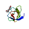 7nerS S: Starting model for refinement |
|---|---|
| Similar structure data |
-Links
 PDBj
PDBj- Assembly
| Deposited unit | 
| ||||||||
|---|---|---|---|---|---|---|---|---|---|
| 1 | 
| ||||||||
| 2 | 
| ||||||||
| Unit cell |
|
-Components
| #1: Protein | Mass: 6861.487 Da / Num. of mol.: 2 / Mutation: Q128R Source method: isolated from a genetically manipulated source Source: (gene. exp.) Rous sarcoma virus (strain Schmidt-Ruppin E)Strain: Schmidt-Ruppin E / Gene: V-SRC / Plasmid: pHTP1 / Production host:  References: UniProt: P63185, non-specific protein-tyrosine kinase #2: Protein/peptide | Mass: 1317.622 Da / Num. of mol.: 2 / Source method: obtained synthetically / Source: (synth.) unidentified (others) #3: Chemical | ChemComp-PEG / |   Mass: 106.120 Da / Num. of mol.: 1 / Source method: obtained synthetically / Formula: C4H10O3 Mass: 106.120 Da / Num. of mol.: 1 / Source method: obtained synthetically / Formula: C4H10O3#4: Chemical | ChemComp-GOL / |   Mass: 92.094 Da / Num. of mol.: 1 / Source method: obtained synthetically / Formula: C3H8O3 Mass: 92.094 Da / Num. of mol.: 1 / Source method: obtained synthetically / Formula: C3H8O3#5: Water | ChemComp-HOH / |  Mass: 18.015 Da / Num. of mol.: 56 / Source method: isolated from a natural source / Formula: H2O Mass: 18.015 Da / Num. of mol.: 56 / Source method: isolated from a natural source / Formula: H2OHas ligand of interest | N | |
|---|
-Experimental details
-Experiment
| Experiment | Method: X-RAY DIFFRACTION / Number of used crystals: 1 |
|---|
- Sample preparation
| Crystal | Density Matthews: 2.15 Å3/Da / Density % sol: 42.68 % / Mosaicity: 0 ° |
|---|---|
| Crystal grow | Temperature: 283 K / Method: vapor diffusion, sitting drop / pH: 6 Details: 2.4 M ammonium sulfate, 10% glicerol, 5% PEG300, 40 mM LiCl, 0.1M MES |
-Data collection
| Diffraction | Mean temperature: 100 K / Serial crystal experiment: N | ||||||||||||||||||||||||||||||
|---|---|---|---|---|---|---|---|---|---|---|---|---|---|---|---|---|---|---|---|---|---|---|---|---|---|---|---|---|---|---|---|
| Diffraction source | Source: SYNCHROTRON / Site: ESRF  / Beamline: MASSIF-3 / Wavelength: 0.9677 Å / Beamline: MASSIF-3 / Wavelength: 0.9677 Å | ||||||||||||||||||||||||||||||
| Detector | Type: DECTRIS EIGER X 4M / Detector: PIXEL / Date: Jun 16, 2021 | ||||||||||||||||||||||||||||||
| Radiation | Protocol: SINGLE WAVELENGTH / Monochromatic (M) / Laue (L): M / Scattering type: x-ray | ||||||||||||||||||||||||||||||
| Radiation wavelength | Wavelength: 0.9677 Å / Relative weight: 1 | ||||||||||||||||||||||||||||||
| Reflection | Resolution: 1.6→17.18 Å / Num. obs: 17900 / % possible obs: 99.6 % / Redundancy: 6.5 % / Biso Wilson estimate: 25.13 Å2 / CC1/2: 0.998 / Rmerge(I) obs: 0.063 / Rpim(I) all: 0.026 / Rrim(I) all: 0.069 / Net I/σ(I): 14.2 / Num. measured all: 116716 / Scaling rejects: 242 | ||||||||||||||||||||||||||||||
| Reflection shell | Diffraction-ID: 1
|
-Phasing
| Phasing | Method: molecular replacement |
|---|
- Processing
| Software |
| ||||||||||||||||||||||||||||||||||||||||||||||||||||||||||||||||||||||||||||||||||||||||||||||||||
|---|---|---|---|---|---|---|---|---|---|---|---|---|---|---|---|---|---|---|---|---|---|---|---|---|---|---|---|---|---|---|---|---|---|---|---|---|---|---|---|---|---|---|---|---|---|---|---|---|---|---|---|---|---|---|---|---|---|---|---|---|---|---|---|---|---|---|---|---|---|---|---|---|---|---|---|---|---|---|---|---|---|---|---|---|---|---|---|---|---|---|---|---|---|---|---|---|---|---|---|
| Refinement | Method to determine structure: MOLECULAR REPLACEMENT Starting model: 7NER Resolution: 1.6→17.18 Å / SU ML: 0.14 / Cross valid method: THROUGHOUT / σ(F): 0.14 / Phase error: 28.03 / Stereochemistry target values: ML
| ||||||||||||||||||||||||||||||||||||||||||||||||||||||||||||||||||||||||||||||||||||||||||||||||||
| Solvent computation | Shrinkage radii: 0.9 Å / VDW probe radii: 1.11 Å / Solvent model: FLAT BULK SOLVENT MODEL | ||||||||||||||||||||||||||||||||||||||||||||||||||||||||||||||||||||||||||||||||||||||||||||||||||
| Displacement parameters | Biso max: 87.97 Å2 / Biso mean: 36.7025 Å2 / Biso min: 17.55 Å2 | ||||||||||||||||||||||||||||||||||||||||||||||||||||||||||||||||||||||||||||||||||||||||||||||||||
| Refinement step | Cycle: final / Resolution: 1.6→17.18 Å
| ||||||||||||||||||||||||||||||||||||||||||||||||||||||||||||||||||||||||||||||||||||||||||||||||||
| LS refinement shell | Refine-ID: X-RAY DIFFRACTION / Rfactor Rfree error: 0 / Total num. of bins used: 13
|
