 Movie
Movie Controller

[English] 日本語
 Yorodumi
- PDB-7pu6: STRUCTURE OF ESTER-HYDROLASE EH7 FROM THE METAGENOME OF MARINE SE... -
+ Open data

- Basic information
| Entry | Database: PDB / ID: 7pu6 | ||||||
|---|---|---|---|---|---|---|---|
| Title | STRUCTURE OF ESTER-HYDROLASE EH7 FROM THE METAGENOME OF MARINE SEDIMENTS AT MILAZZO HARBOR (SICILY, ITALY) COMPLEXED WITH A DERIVATIVE OF OCTYL 4-NITROPHENYL HEXYLPHOSPHONATE | ||||||
 Components Components | Esterase | ||||||
 Keywords Keywords | HYDROLASE / Ester Hydrolase / Complex | ||||||
| Function / homology | : / Beta-lactamase-related / Beta-lactamase / Beta-lactamase/transpeptidase-like / hexyl(octoxy)phosphinic acid / Esterase Function and homology information Function and homology information | ||||||
| Biological species | metagenome (others) | ||||||
| Method |  X-RAY DIFFRACTION / SYNCHROTRON / MOLECULAR REPLACEMENT / Resolution: 2.92 Å X-RAY DIFFRACTION / SYNCHROTRON / MOLECULAR REPLACEMENT / Resolution: 2.92 Å | ||||||
 Authors Authors | Cea Rama, I. / Sanz-Aparicio, J. | ||||||
| Funding support |  Spain, 1items Spain, 1items
| ||||||
 Citation Citation | Journal: Febs J. / Year: 2022 Title: Crystal structure of a family VIII beta-lactamase fold hydrolase reveals the molecular mechanism for its broad substrate scope. Authors: Cea-Rama, I. / Coscolin, C. / Gonzalez-Alfonso, J.L. / Raj, J. / Vasiljevic, M. / Plou, F.J. / Ferrer, M. / Sanz-Aparicio, J. | ||||||
| History |
|
- Structure visualization
| Structure viewer | Molecule: MolmilJmol/JSmol |
|---|
- Downloads & links
-Download
| PDBx/mmCIF format | 7pu6.cif.gz | 613.8 KB | Display | PDBx/mmCIF format |
|---|---|---|---|---|
| PDB format | pdb7pu6.ent.gz | 507.5 KB | Display | PDB format |
| PDBx/mmJSON format | 7pu6.json.gz | Tree view | PDBx/mmJSON format | |
| Others |  Other downloads Other downloads |
-Validation report
| Arichive directory | https://data.pdbj.org/pub/pdb/validation_reports/pu/7pu6ftp://data.pdbj.org/pub/pdb/validation_reports/pu/7pu6 | HTTPS FTP |
|---|
-Related structure data
| Related structure data |  7pp3SC  7pp8C S: Starting model for refinement C: citing same article ( |
|---|---|
| Similar structure data |
-Links
 PDBj
PDBj
- Assembly
| Deposited unit | 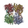
| |||||||||||||||||||||||||||||||||||||||||||||||||||||||||||||||||||||||||||||||||||||||||||||||||||||||||||||||||||||||||||||||||||||||||||||||||||||||||||||||||||||||||||||||||||||||||||||||||||||||||||||||||||||||||||||||||||||||||||||||||||||||||||||||||||||||||||||||||||||||||||||||||||||||||||||||||||||||||||
|---|---|---|---|---|---|---|---|---|---|---|---|---|---|---|---|---|---|---|---|---|---|---|---|---|---|---|---|---|---|---|---|---|---|---|---|---|---|---|---|---|---|---|---|---|---|---|---|---|---|---|---|---|---|---|---|---|---|---|---|---|---|---|---|---|---|---|---|---|---|---|---|---|---|---|---|---|---|---|---|---|---|---|---|---|---|---|---|---|---|---|---|---|---|---|---|---|---|---|---|---|---|---|---|---|---|---|---|---|---|---|---|---|---|---|---|---|---|---|---|---|---|---|---|---|---|---|---|---|---|---|---|---|---|---|---|---|---|---|---|---|---|---|---|---|---|---|---|---|---|---|---|---|---|---|---|---|---|---|---|---|---|---|---|---|---|---|---|---|---|---|---|---|---|---|---|---|---|---|---|---|---|---|---|---|---|---|---|---|---|---|---|---|---|---|---|---|---|---|---|---|---|---|---|---|---|---|---|---|---|---|---|---|---|---|---|---|---|---|---|---|---|---|---|---|---|---|---|---|---|---|---|---|---|---|---|---|---|---|---|---|---|---|---|---|---|---|---|---|---|---|---|---|---|---|---|---|---|---|---|---|---|---|---|---|---|---|---|---|---|---|---|---|---|---|---|---|---|---|---|---|---|---|---|---|---|---|---|---|---|---|---|---|---|---|---|---|---|---|---|---|---|---|---|---|---|---|---|---|---|---|---|---|---|---|---|---|
| 1 |
| |||||||||||||||||||||||||||||||||||||||||||||||||||||||||||||||||||||||||||||||||||||||||||||||||||||||||||||||||||||||||||||||||||||||||||||||||||||||||||||||||||||||||||||||||||||||||||||||||||||||||||||||||||||||||||||||||||||||||||||||||||||||||||||||||||||||||||||||||||||||||||||||||||||||||||||||||||||||||||
| Unit cell |
| |||||||||||||||||||||||||||||||||||||||||||||||||||||||||||||||||||||||||||||||||||||||||||||||||||||||||||||||||||||||||||||||||||||||||||||||||||||||||||||||||||||||||||||||||||||||||||||||||||||||||||||||||||||||||||||||||||||||||||||||||||||||||||||||||||||||||||||||||||||||||||||||||||||||||||||||||||||||||||
| Noncrystallographic symmetry (NCS) | NCS domain:
NCS domain segments: Component-ID: _ / Beg auth comp-ID: SER / Beg label comp-ID: SER / End auth comp-ID: ASP / End label comp-ID: ASP / Refine code: _ / Auth seq-ID: 8 - 409 / Label seq-ID: 27 - 428
|