[English] 日本語
Yorodumi
- PDB-7ptb: Crystal structure of the SPD-2 domain of human CEP192 -

+
Open data


ID or keywords:

Loading...

-
Basic information

Entry
Database: PDB / ID: 7ptb
TitleCrystal structure of the SPD-2 domain of human CEP192
ComponentsCentrosomal protein of 192 kDaCentrosome
KeywordsCELL CYCLE / Centriole / Centrosome / Cell-cycle / Pericentriolar material
Function / homology
Function and homology information


centrosome-templated microtubule nucleation / procentriole / procentriole replication complex / protein localization to centrosome / pericentriolar material / centriole replication / mitotic spindle assembly / phosphatase binding / Loss of Nlp from mitotic centrosomes / Loss of proteins required for interphase microtubule organization from the centrosome ...centrosome-templated microtubule nucleation / procentriole / procentriole replication complex / protein localization to centrosome / pericentriolar material / centriole replication / mitotic spindle assembly / phosphatase binding / Loss of Nlp from mitotic centrosomes / Loss of proteins required for interphase microtubule organization from the centrosome / Recruitment of mitotic centrosome proteins and complexes / Recruitment of NuMA to mitotic centrosomes / Anchoring of the basal body to the plasma membrane / centriole / AURKA Activation by TPX2 / response to bacterium / Regulation of PLK1 Activity at G2/M Transition / centrosome / protein kinase binding / cytosol / cytoplasm
Similarity search - Function
Centrosomal protein Spd-2/CEP192 / Immunoglobulin-like fold
Similarity search - Domain/homology
NITRATE ION / Centrosomal protein of 192 kDa
Similarity search - Component
Biological speciesHomo sapiens (human)
MethodX-RAY DIFFRACTION / SYNCHROTRON / SAD / Resolution: 2.08 Å
AuthorsRosa e Silva, I. / van Breugel, M.
Funding support United Kingdom, 1items
OrganizationGrant numberCountry
Medical Research Council (MRC, United Kingdom)MRC - MC_UP_1201/3 United Kingdom
CitationJournal: Commun Biol / Year: 2022
Title: Structural validation and assessment of AlphaFold2 predictions for centrosomal and centriolar proteins and their complexes.
Authors: van Breugel, M. / Rosa E Silva, I. / Andreeva, A.
History
DepositionSep 26, 2021Deposition site: PDBE / Processing site: PDBE
Revision 1.0Apr 6, 2022Provider: repository / Type: Initial release
Revision 1.1Apr 20, 2022Group: Database references / Category: citation / citation_author
Item: _citation.journal_volume / _citation.page_first ..._citation.journal_volume / _citation.page_first / _citation.page_last / _citation.pdbx_database_id_PubMed / _citation_author.identifier_ORCID / _citation_author.name

-
Structure visualization

Structure viewerMolecule:
MolmilJmol/JSmol

Downloads & links

-
Assembly

Deposited unit
A: Centrosomal protein of 192 kDa
hetero molecules


Theoretical massNumber of molelcules
Total (without water)39,5274
Polymers39,3411
Non-polymers1863
Water2,882160
1


  • Idetical with deposited unit
  • defined by author&software
  • Evidence: light scattering, SEC MALS suggests a monomeric protein
TypeNameSymmetry operationNumber
identity operation1_555x,y,z1
Buried area450 Å2
ΔGint3 kcal/mol
Surface area17790 Å2
MethodPISA
Unit cell
Length a, b, c (Å)104.588, 104.588, 90.214
Angle α, β, γ (deg.)90.00, 90.00, 120.00
Int Tables number169
Space group name H-MP61

-
Components

#1: Protein Centrosomal protein of 192 kDa / Centrosome / Cep192 / Cep192/SPD-2


Mass: 39340.531 Da / Num. of mol.: 1
Source method: isolated from a genetically manipulated source
Details: The GP residues on the N-terminus were left after PreScission protease cleavage.
Source: (gene. exp.) Homo sapiens (human) / Gene: CEP192, KIAA1569, PP8407 / Cell line (production host): Sf9 / Production host: 
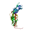
Spodoptera frugiperda (fall armyworm) / References: UniProt: Q8TEP8
#2: Chemical ChemComp-NO3 / NITRATE ION / Nitrate


Mass: 62.005 Da / Num. of mol.: 3 / Source method: obtained synthetically / Formula: NO3
#3: Water ChemComp-HOH / water / Water


Mass: 18.015 Da / Num. of mol.: 160 / Source method: isolated from a natural source / Formula: H2O
Has ligand of interestN

-
Experimental details

-
Experiment

ExperimentMethod: X-RAY DIFFRACTION / Number of used crystals: 1

-
Sample preparation

CrystalDensity Matthews: 3.64 Å3/Da / Density % sol: 66.19 %
Crystal growTemperature: 292.15 K / Method: vapor diffusion
Details: 100 nl protein solution and 100 nl of reservoir solution which was 0.1 M Na-Acetate pH 5.3, 3.9 M ammonium ni-trate. Crystals were mounted in 0.1 M Na-Acetate pH 4.6, 1 M ammonium nitrate, 30% glycerol.

-
Data collection

DiffractionMean temperature: 80 K / Serial crystal experiment: N
Diffraction sourceSource: SYNCHROTRON / Site: Diamond / Beamline: I24 / Wavelength: 0.97835 Å
DetectorType: DECTRIS PILATUS3 6M / Detector: PIXEL / Date: May 9, 2018
RadiationProtocol: SINGLE WAVELENGTH / Monochromatic (M) / Laue (L): M / Scattering type: x-ray
Radiation wavelengthWavelength: 0.97835 Å / Relative weight: 1
ReflectionResolution: 2.08→90.6 Å / Num. obs: 33669 / % possible obs: 100 % / Redundancy: 18.6 % / Rpim(I) all: 0.024 / Net I/σ(I): 19.7
Reflection shellResolution: 2.08→2.14 Å / Redundancy: 2.5 % / Num. unique obs: 32002 / Rpim(I) all: 0.357 / % possible all: 100

-
Processing

Software
NameVersionClassification
REFMAC5.8.0267refinement
PHENIX1.18refinement
iMOSFLMdata reduction
Aimlessdata scaling
CRANK2phasing
RefinementMethod to determine structure: SAD / Resolution: 2.08→90.58 Å / Cor.coef. Fo:Fc: 0.948 / Cor.coef. Fo:Fc free: 0.946 / SU B: 4.086 / SU ML: 0.105 / Cross valid method: THROUGHOUT / ESU R: 0.162 / ESU R Free: 0.138 / Stereochemistry target values: MAXIMUM LIKELIHOOD / Details: HYDROGENS HAVE BEEN ADDED IN THE RIDING POSITIONS
RfactorNum. reflection% reflectionSelection details
Rfree0.22364 1629 4.8 %RANDOM
Rwork0.21197 ---
obs0.21252 32002 99.99 %-
Solvent computationIon probe radii: 0.8 Å / Shrinkage radii: 0.8 Å / VDW probe radii: 1.2 Å / Solvent model: MASK
Displacement parametersBiso mean: 45.671 Å2
Baniso -1Baniso -2Baniso -3
1-0.32 Å20.16 Å20 Å2
2--0.32 Å2-0 Å2
3----1.03 Å2
Refinement stepCycle: 1 / Resolution: 2.08→90.58 Å
ProteinNucleic acidLigandSolventTotal
Num. atoms2516 0 12 160 2688
Refine LS restraints
Refine-IDTypeDev idealDev ideal targetNumber
X-RAY DIFFRACTIONr_bond_refined_d0.0020.0132626
X-RAY DIFFRACTIONr_bond_other_d0.0010.0172594
X-RAY DIFFRACTIONr_angle_refined_deg1.1711.6483553
X-RAY DIFFRACTIONr_angle_other_deg1.0291.5795979
X-RAY DIFFRACTIONr_dihedral_angle_1_deg6.7025334
X-RAY DIFFRACTIONr_dihedral_angle_2_deg32.09721.857140
X-RAY DIFFRACTIONr_dihedral_angle_3_deg12.00915481
X-RAY DIFFRACTIONr_dihedral_angle_4_deg13.4371521
X-RAY DIFFRACTIONr_chiral_restr0.0360.2346
X-RAY DIFFRACTIONr_gen_planes_refined0.0020.022969
X-RAY DIFFRACTIONr_gen_planes_other0.0010.02621
X-RAY DIFFRACTIONr_nbd_refined
X-RAY DIFFRACTIONr_nbd_other
X-RAY DIFFRACTIONr_nbtor_refined
X-RAY DIFFRACTIONr_nbtor_other
X-RAY DIFFRACTIONr_xyhbond_nbd_refined
X-RAY DIFFRACTIONr_xyhbond_nbd_other
X-RAY DIFFRACTIONr_metal_ion_refined
X-RAY DIFFRACTIONr_metal_ion_other
X-RAY DIFFRACTIONr_symmetry_vdw_refined
X-RAY DIFFRACTIONr_symmetry_vdw_other
X-RAY DIFFRACTIONr_symmetry_hbond_refined
X-RAY DIFFRACTIONr_symmetry_hbond_other
X-RAY DIFFRACTIONr_symmetry_metal_ion_refined
X-RAY DIFFRACTIONr_symmetry_metal_ion_other
X-RAY DIFFRACTIONr_mcbond_it1.2634.7761285
X-RAY DIFFRACTIONr_mcbond_other1.2644.7731284
X-RAY DIFFRACTIONr_mcangle_it2.3437.1551609
X-RAY DIFFRACTIONr_mcangle_other2.3437.1581610
X-RAY DIFFRACTIONr_scbond_it0.94.9491341
X-RAY DIFFRACTIONr_scbond_other0.8984.9481333
X-RAY DIFFRACTIONr_scangle_it
X-RAY DIFFRACTIONr_scangle_other1.6737.3581927
X-RAY DIFFRACTIONr_long_range_B_refined4.55253.6172654
X-RAY DIFFRACTIONr_long_range_B_other4.36753.4262624
X-RAY DIFFRACTIONr_rigid_bond_restr
X-RAY DIFFRACTIONr_sphericity_free
X-RAY DIFFRACTIONr_sphericity_bonded
LS refinement shellResolution: 2.082→2.136 Å / Total num. of bins used: 20
RfactorNum. reflection% reflection
Rfree0.311 103 -
Rwork0.287 2368 -
obs--99.92 %

+
About Yorodumi

-
News

-
Feb 9, 2022. New format data for meta-information of EMDB entries

New format data for meta-information of EMDB entries

  • Version 3 of the EMDB header file is now the official format.
  • The previous official version 1.9 will be removed from the archive.

Related info.:EMDB header

External links:wwPDB to switch to version 3 of the EMDB data model

-
Aug 12, 2020. Covid-19 info

Covid-19 info

URL: https://pdbj.org/emnavi/covid19.php

New page: Covid-19 featured information page in EM Navigator.

Related info.:Covid-19 info / Mar 5, 2020. Novel coronavirus structure data

+
Mar 5, 2020. Novel coronavirus structure data

Novel coronavirus structure data

Related info.:Yorodumi Speices / Aug 12, 2020. Covid-19 info

External links:COVID-19 featured content - PDBj / Molecule of the Month (242):Coronavirus Proteases

+
Jan 31, 2019. EMDB accession codes are about to change! (news from PDBe EMDB page)

EMDB accession codes are about to change! (news from PDBe EMDB page)

  • The allocation of 4 digits for EMDB accession codes will soon come to an end. Whilst these codes will remain in use, new EMDB accession codes will include an additional digit and will expand incrementally as the available range of codes is exhausted. The current 4-digit format prefixed with “EMD-” (i.e. EMD-XXXX) will advance to a 5-digit format (i.e. EMD-XXXXX), and so on. It is currently estimated that the 4-digit codes will be depleted around Spring 2019, at which point the 5-digit format will come into force.
  • The EM Navigator/Yorodumi systems omit the EMD- prefix.

Related info.:Q: What is EMD? / ID/Accession-code notation in Yorodumi/EM Navigator

External links:EMDB Accession Codes are Changing Soon! / Contact to PDBj

+
Jul 12, 2017. Major update of PDB

Major update of PDB

  • wwPDB released updated PDB data conforming to the new PDBx/mmCIF dictionary.
  • This is a major update changing the version number from 4 to 5, and with Remediation, in which all the entries are updated.
  • In this update, many items about electron microscopy experimental information are reorganized (e.g. em_software).
  • Now, EM Navigator and Yorodumi are based on the updated data.

External links:wwPDB Remediation / Enriched Model Files Conforming to OneDep Data Standards Now Available in the PDB FTP Archive

-
Yorodumi

Thousand views of thousand structures

  • Yorodumi is a browser for structure data from EMDB, PDB, SASBDB, etc.
  • This page is also the successor to EM Navigator detail page, and also detail information page/front-end page for Omokage search.
  • The word "yorodu" (or yorozu) is an old Japanese word meaning "ten thousand". "mi" (miru) is to see.

Related info.:EMDB / PDB / SASBDB / Comparison of 3 databanks / Yorodumi Search / Aug 31, 2016. New EM Navigator & Yorodumi / Yorodumi Papers / Jmol/JSmol / Function and homology information / Changes in new EM Navigator and Yorodumi

Read more