 Movie
Movie Controller

[English] 日本語
 Yorodumi
- PDB-7psw: Spin labeled IPNS S55C variant in complex with Fe and ACV under a... -
+ Open data

- Basic information
| Entry | Database: PDB / ID: 7psw | ||||||
|---|---|---|---|---|---|---|---|
| Title | Spin labeled IPNS S55C variant in complex with Fe and ACV under anaerobic conditions | ||||||
 Components Components | Isopenicillin N synthase | ||||||
 Keywords Keywords | OXIDOREDUCTASE / Isopenicillin N synthase / Oxidase / Penicillin biosynthesis / Spin labeled protein | ||||||
| Function / homology |  Function and homology information Function and homology informationisopenicillin-N synthase / isopenicillin-N synthase activity / penicillin biosynthetic process / L-ascorbic acid binding / iron ion binding / cytosol Similarity search - Function | ||||||
| Biological species |  | ||||||
| Method |  X-RAY DIFFRACTION / SYNCHROTRON / MOLECULAR REPLACEMENT / Resolution: 1.21 Å X-RAY DIFFRACTION / SYNCHROTRON / MOLECULAR REPLACEMENT / Resolution: 1.21 Å | ||||||
 Authors Authors | Rabe, P. / Clifton, I. / Walla, C. / Schofield, C.J. | ||||||
| Funding support |  United Kingdom, 1items United Kingdom, 1items
| ||||||
 Citation Citation | Journal: J.Biol.Chem. / Year: 2022 Title: Spectroscopic studies reveal details of substrate-induced conformational changes distant from the active site in isopenicillin N synthase. Authors: Rabe, P. / Walla, C.C. / Goodyear, N.K. / Welsh, J. / Southwart, R. / Clifton, I. / Linyard, J.D.S. / Tumber, A. / Claridge, T.D.W. / Myers, W.K. / Schofield, C.J. | ||||||
| History |
|
- Structure visualization
| Structure viewer | Molecule: MolmilJmol/JSmol |
|---|
- Downloads & links
-Download
| PDBx/mmCIF format | 7psw.cif.gz | 228.2 KB | Display | PDBx/mmCIF format |
|---|---|---|---|---|
| PDB format | pdb7psw.ent.gz | 185.1 KB | Display | PDB format |
| PDBx/mmJSON format | 7psw.json.gz | Tree view | PDBx/mmJSON format | |
| Others |  Other downloads Other downloads |
-Validation report
| Arichive directory | https://data.pdbj.org/pub/pdb/validation_reports/ps/7pswftp://data.pdbj.org/pub/pdb/validation_reports/ps/7psw | HTTPS FTP |
|---|
-Related structure data
| Related structure data |  7poyC 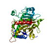 6zaeS C: citing same article ( S: Starting model for refinement |
|---|---|
| Similar structure data |

-Links
 PDBj
PDBj
- Assembly
| Deposited unit | 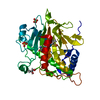
| ||||||||
|---|---|---|---|---|---|---|---|---|---|
| 1 |
| ||||||||
| Unit cell |
|
-Components
-Protein , 1 types, 1 molecules A
| #1: Protein | Mass: 37579.902 Da / Num. of mol.: 1 / Mutation: Ser55Cys Source method: isolated from a genetically manipulated source Source: (gene. exp.) Strain: FGSC A4 / ATCC 38163 / CBS 112.46 / NRRL 194 / M139 / Gene: ipnA, ips, AN2622 / Plasmid: pCOLD_IPNS / Details (production host): pCOLD_IPNS_S55C / Production host:  |
|---|
-Non-polymers , 5 types, 470 molecules 


| #2: Chemical | ChemComp-FE /  Mass: 55.845 Da / Num. of mol.: 1 / Source method: obtained synthetically / Formula: Fe / Feature type: SUBJECT OF INVESTIGATION Mass: 55.845 Da / Num. of mol.: 1 / Source method: obtained synthetically / Formula: Fe / Feature type: SUBJECT OF INVESTIGATION | ||||
|---|---|---|---|---|---|
| #3: Chemical | ChemComp-ACV /  Mass: 363.430 Da / Num. of mol.: 1 / Source method: obtained synthetically / Formula: C14H25N3O6S / Feature type: SUBJECT OF INVESTIGATION Mass: 363.430 Da / Num. of mol.: 1 / Source method: obtained synthetically / Formula: C14H25N3O6S / Feature type: SUBJECT OF INVESTIGATION | ||||
| #4: Chemical |  Mass: 96.063 Da / Num. of mol.: 3 / Source method: obtained synthetically / Formula: SO4 Mass: 96.063 Da / Num. of mol.: 3 / Source method: obtained synthetically / Formula: SO4#5: Chemical | ChemComp-GOL / |  Mass: 92.094 Da / Num. of mol.: 1 / Source method: obtained synthetically / Formula: C3H8O3 Mass: 92.094 Da / Num. of mol.: 1 / Source method: obtained synthetically / Formula: C3H8O3#6: Water | ChemComp-HOH / | Mass: 18.015 Da / Num. of mol.: 464 / Source method: isolated from a natural source / Formula: H2O |
-Details
| Has ligand of interest | Y |
|---|
-Experimental details
-Experiment
| Experiment | Method: X-RAY DIFFRACTION / Number of used crystals: 1 |
|---|
- Sample preparation
| Crystal | Density Matthews: 2.24 Å3/Da / Density % sol: 45.12 % / Description: plate morphology, 50 x 200 um |
|---|---|
| Crystal grow | Temperature: 298 K / Method: vapor diffusion, hanging drop / pH: 8.5 / Details: 1.7 M Li2SO4, 0.1 M Tris pH 8.5 |
-Data collection
| Diffraction | Mean temperature: 100 K / Ambient temp details: cryo / Serial crystal experiment: N | |||||||||||||||||||||||||||
|---|---|---|---|---|---|---|---|---|---|---|---|---|---|---|---|---|---|---|---|---|---|---|---|---|---|---|---|---|
| Diffraction source | Source: SYNCHROTRON / Site: Diamond / Beamline: I03 / Wavelength: 0.9763 Å | |||||||||||||||||||||||||||
| Detector | Type: DECTRIS EIGER2 XE 16M / Detector: PIXEL / Date: Jan 21, 2021 | |||||||||||||||||||||||||||
| Radiation | Protocol: SINGLE WAVELENGTH / Monochromatic (M) / Laue (L): M / Scattering type: x-ray | |||||||||||||||||||||||||||
| Radiation wavelength | Wavelength: 0.9763 Å / Relative weight: 1 | |||||||||||||||||||||||||||
| Reflection | Resolution: 1.21→58.35 Å / Num. obs: 103529 / % possible obs: 99.9 % / Redundancy: 12.2 % / Biso Wilson estimate: 11.96 Å2 / CC1/2: 1 / Rmerge(I) obs: 0.05 / Rpim(I) all: 0.015 / Rrim(I) all: 0.052 / Net I/σ(I): 24.2 | |||||||||||||||||||||||||||
| Reflection shell | Diffraction-ID: 1
|
- Processing
| Software |
| |||||||||||||||||||||||||||||||||||||||||||||||||||||||||||||||||||||||||||||||||||||||||||||||||||||||||||||||||||||||||||||||||||||||||||||||||||||||||||||||||||||||||||||||||||||||||||||||||||||||||||||||||||||||||
|---|---|---|---|---|---|---|---|---|---|---|---|---|---|---|---|---|---|---|---|---|---|---|---|---|---|---|---|---|---|---|---|---|---|---|---|---|---|---|---|---|---|---|---|---|---|---|---|---|---|---|---|---|---|---|---|---|---|---|---|---|---|---|---|---|---|---|---|---|---|---|---|---|---|---|---|---|---|---|---|---|---|---|---|---|---|---|---|---|---|---|---|---|---|---|---|---|---|---|---|---|---|---|---|---|---|---|---|---|---|---|---|---|---|---|---|---|---|---|---|---|---|---|---|---|---|---|---|---|---|---|---|---|---|---|---|---|---|---|---|---|---|---|---|---|---|---|---|---|---|---|---|---|---|---|---|---|---|---|---|---|---|---|---|---|---|---|---|---|---|---|---|---|---|---|---|---|---|---|---|---|---|---|---|---|---|---|---|---|---|---|---|---|---|---|---|---|---|---|---|---|---|---|---|---|---|---|---|---|---|---|---|---|---|---|---|---|---|---|
| Refinement | Method to determine structure: MOLECULAR REPLACEMENT Starting model: 6ZAE Resolution: 1.21→41.26 Å / SU ML: 0.11 / Cross valid method: THROUGHOUT / σ(F): 1.36 / Phase error: 15.7 / Stereochemistry target values: ML
| |||||||||||||||||||||||||||||||||||||||||||||||||||||||||||||||||||||||||||||||||||||||||||||||||||||||||||||||||||||||||||||||||||||||||||||||||||||||||||||||||||||||||||||||||||||||||||||||||||||||||||||||||||||||||
| Solvent computation | Shrinkage radii: 0.9 Å / VDW probe radii: 1.11 Å / Solvent model: FLAT BULK SOLVENT MODEL | |||||||||||||||||||||||||||||||||||||||||||||||||||||||||||||||||||||||||||||||||||||||||||||||||||||||||||||||||||||||||||||||||||||||||||||||||||||||||||||||||||||||||||||||||||||||||||||||||||||||||||||||||||||||||
| Displacement parameters | Biso max: 105.97 Å2 / Biso mean: 18.4682 Å2 / Biso min: 6.94 Å2 | |||||||||||||||||||||||||||||||||||||||||||||||||||||||||||||||||||||||||||||||||||||||||||||||||||||||||||||||||||||||||||||||||||||||||||||||||||||||||||||||||||||||||||||||||||||||||||||||||||||||||||||||||||||||||
| Refinement step | Cycle: final / Resolution: 1.21→41.26 Å
| |||||||||||||||||||||||||||||||||||||||||||||||||||||||||||||||||||||||||||||||||||||||||||||||||||||||||||||||||||||||||||||||||||||||||||||||||||||||||||||||||||||||||||||||||||||||||||||||||||||||||||||||||||||||||
| LS refinement shell | Refine-ID: X-RAY DIFFRACTION / Rfactor Rfree error: 0 / Total num. of bins used: 30
| |||||||||||||||||||||||||||||||||||||||||||||||||||||||||||||||||||||||||||||||||||||||||||||||||||||||||||||||||||||||||||||||||||||||||||||||||||||||||||||||||||||||||||||||||||||||||||||||||||||||||||||||||||||||||
| Refinement TLS params. | Method: refined / Refine-ID: X-RAY DIFFRACTION
| |||||||||||||||||||||||||||||||||||||||||||||||||||||||||||||||||||||||||||||||||||||||||||||||||||||||||||||||||||||||||||||||||||||||||||||||||||||||||||||||||||||||||||||||||||||||||||||||||||||||||||||||||||||||||
| Refinement TLS group |
|
