[English] 日本語
 Yorodumi
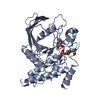
- PDB-7ppn: SHP2 catalytic domain in complex with CD28 (183-198) phosphopepti... -
+ Open data
- Basic information
| Entry | Database: PDB / ID: 7ppn | |||||||||
|---|---|---|---|---|---|---|---|---|---|---|
| Title | SHP2 catalytic domain in complex with CD28 (183-198) phosphopeptide (pTyr-191, p-Thr-195) | |||||||||
 Components Components |
| |||||||||
 Keywords Keywords | SIGNALING PROTEIN / SHP2 / Phosphatase / CD28 / phosphopeptide / signaling | |||||||||
| Function / homology |  Function and homology information Function and homology informationNef mediated downregulation of CD28 cell surface expression / positive regulation of inflammatory response to antigenic stimulus / regulatory T cell differentiation / protein complex involved in cell adhesion / negative regulation of cortisol secretion / intestinal epithelial cell migration / microvillus organization / negative regulation of growth hormone secretion / genitalia development / atrioventricular canal development ...Nef mediated downregulation of CD28 cell surface expression / positive regulation of inflammatory response to antigenic stimulus / regulatory T cell differentiation / protein complex involved in cell adhesion / negative regulation of cortisol secretion / intestinal epithelial cell migration / microvillus organization / negative regulation of growth hormone secretion / genitalia development / atrioventricular canal development / regulation of regulatory T cell differentiation / negative regulation of cell adhesion mediated by integrin / positive regulation of isotype switching to IgG isotypes / CD4-positive, alpha-beta T cell proliferation / STAT5 Activation / Co-inhibition by BTLA / negative thymic T cell selection / Netrin mediated repulsion signals / cerebellar cortex formation / negative regulation of neutrophil activation / positive regulation of CD4-positive, alpha-beta T cell proliferation / positive regulation of hormone secretion / regulation of protein export from nucleus / positive regulation of ossification / positive regulation of lipopolysaccharide-mediated signaling pathway / Interleukin-37 signaling / Signaling by Leptin / hormone metabolic process / MET activates PTPN11 / Co-stimulation by CD28 / Regulation of RUNX1 Expression and Activity / negative regulation of chondrocyte differentiation / CD28 dependent Vav1 pathway / Signal regulatory protein family interactions / face morphogenesis / ERBB signaling pathway / platelet formation / triglyceride metabolic process / megakaryocyte development / organ growth / negative regulation of type I interferon production / Interleukin-20 family signaling / PI-3K cascade:FGFR3 / Interleukin-6 signaling / Co-inhibition by CTLA4 / Platelet sensitization by LDL / STAT5 activation downstream of FLT3 ITD mutants / peptide hormone receptor binding / PI-3K cascade:FGFR2 / PI-3K cascade:FGFR4 / MAPK3 (ERK1) activation / PI-3K cascade:FGFR1 / Prolactin receptor signaling / neurotrophin TRK receptor signaling pathway / positive regulation of interleukin-4 production / regulation of cell adhesion mediated by integrin / regulation of type I interferon-mediated signaling pathway / platelet-derived growth factor receptor signaling pathway / MAPK1 (ERK2) activation / PECAM1 interactions / inner ear development / Bergmann glial cell differentiation / peptidyl-tyrosine dephosphorylation / positive regulation of intracellular signal transduction / humoral immune response / non-membrane spanning protein tyrosine phosphatase activity / phosphoprotein phosphatase activity / positive regulation of interleukin-10 production / RET signaling / Regulation of IFNA/IFNB signaling / Interleukin-3, Interleukin-5 and GM-CSF signaling / PI3K Cascade / immunological synapse / Co-inhibition by PD-1 / CD28 dependent PI3K/Akt signaling / fibroblast growth factor receptor signaling pathway / GAB1 signalosome / positive regulation of insulin receptor signaling pathway / ephrin receptor signaling pathway / regulation of protein-containing complex assembly / Activated NTRK2 signals through FRS2 and FRS3 / Regulation of IFNG signaling / positive regulation of viral genome replication / coreceptor activity / cell adhesion molecule binding / FRS-mediated FGFR3 signaling / Signaling by CSF3 (G-CSF) / GPVI-mediated activation cascade / Signaling by FLT3 ITD and TKD mutants / negative regulation of T cell proliferation / T cell costimulation / FRS-mediated FGFR2 signaling / hormone-mediated signaling pathway / FRS-mediated FGFR4 signaling / Activation of IRF3, IRF7 mediated by TBK1, IKKε (IKBKE) / FRS-mediated FGFR1 signaling / Tie2 Signaling / positive regulation of T cell proliferation / phosphotyrosine residue binding / FLT3 Signaling Similarity search - Function | |||||||||
| Biological species |  Homo sapiens (human) Homo sapiens (human) | |||||||||
| Method |  X-RAY DIFFRACTION / X-RAY DIFFRACTION /  SYNCHROTRON / SYNCHROTRON /  MOLECULAR REPLACEMENT / MOLECULAR REPLACEMENT /  molecular replacement / Resolution: 1.9 Å molecular replacement / Resolution: 1.9 Å | |||||||||
 Authors Authors | Sok, P. / Zeke, A. / Remenyi, A. | |||||||||
| Funding support |  Hungary, 2items Hungary, 2items
| |||||||||
 Citation Citation |  Journal: Nat Commun / Year: 2022 Journal: Nat Commun / Year: 2022Title: Structural insights into the pSer/pThr dependent regulation of the SHP2 tyrosine phosphatase in insulin and CD28 signaling. Authors: Zeke, A. / Takacs, T. / Sok, P. / Nemeth, K. / Kirsch, K. / Egri, P. / Poti, A.L. / Bento, I. / Tusnady, G.E. / Remenyi, A. #1: Journal: Acta Crystallogr D Struct Biol / Year: 2019 Title: Macromolecular structure determination using X-rays, neutrons and electrons: recent developments in Phenix. Authors: Dorothee Liebschner / Pavel V Afonine / Matthew L Baker / Gábor Bunkóczi / Vincent B Chen / Tristan I Croll / Bradley Hintze / Li Wei Hung / Swati Jain / Airlie J McCoy / Nigel W Moriarty ...Authors: Dorothee Liebschner / Pavel V Afonine / Matthew L Baker / Gábor Bunkóczi / Vincent B Chen / Tristan I Croll / Bradley Hintze / Li Wei Hung / Swati Jain / Airlie J McCoy / Nigel W Moriarty / Robert D Oeffner / Billy K Poon / Michael G Prisant / Randy J Read / Jane S Richardson / David C Richardson / Massimo D Sammito / Oleg V Sobolev / Duncan H Stockwell / Thomas C Terwilliger / Alexandre G Urzhumtsev / Lizbeth L Videau / Christopher J Williams / Paul D Adams /    Abstract: Diffraction (X-ray, neutron and electron) and electron cryo-microscopy are powerful methods to determine three-dimensional macromolecular structures, which are required to understand biological ...Diffraction (X-ray, neutron and electron) and electron cryo-microscopy are powerful methods to determine three-dimensional macromolecular structures, which are required to understand biological processes and to develop new therapeutics against diseases. The overall structure-solution workflow is similar for these techniques, but nuances exist because the properties of the reduced experimental data are different. Software tools for structure determination should therefore be tailored for each method. Phenix is a comprehensive software package for macromolecular structure determination that handles data from any of these techniques. Tasks performed with Phenix include data-quality assessment, map improvement, model building, the validation/rebuilding/refinement cycle and deposition. Each tool caters to the type of experimental data. The design of Phenix emphasizes the automation of procedures, where possible, to minimize repetitive and time-consuming manual tasks, while default parameters are chosen to encourage best practice. A graphical user interface provides access to many command-line features of Phenix and streamlines the transition between programs, project tracking and re-running of previous tasks. | |||||||||
| History |
|
- Structure visualization
| Structure viewer | Molecule:  Molmil Molmil Jmol/JSmol Jmol/JSmol |
|---|
- Downloads & links
- Download
| PDBx/mmCIF format |  7ppn.cif.gz 7ppn.cif.gz | 142.2 KB | Display |  PDBx/mmCIF format PDBx/mmCIF format |
|---|---|---|---|---|
| PDB format |  pdb7ppn.ent.gz pdb7ppn.ent.gz | 104.5 KB | Display |  PDB format PDB format |
| PDBx/mmJSON format |  7ppn.json.gz 7ppn.json.gz | Tree view |  PDBx/mmJSON format PDBx/mmJSON format | |
| Others |  Other downloads Other downloads |
-Validation report
| Summary document |  7ppn_validation.pdf.gz 7ppn_validation.pdf.gz | 448.4 KB | Display |  wwPDB validaton report wwPDB validaton report |
|---|---|---|---|---|
| Full document |  7ppn_full_validation.pdf.gz 7ppn_full_validation.pdf.gz | 449.3 KB | Display | |
| Data in XML |  7ppn_validation.xml.gz 7ppn_validation.xml.gz | 12.4 KB | Display | |
| Data in CIF |  7ppn_validation.cif.gz 7ppn_validation.cif.gz | 16.6 KB | Display | |
| Arichive directory |  https://data.pdbj.org/pub/pdb/validation_reports/pp/7ppn https://data.pdbj.org/pub/pdb/validation_reports/pp/7ppn ftp://data.pdbj.org/pub/pdb/validation_reports/pp/7ppn ftp://data.pdbj.org/pub/pdb/validation_reports/pp/7ppn | HTTPS FTP |
-Related structure data
| Related structure data |  7pplC  7ppmC  3zm0S S: Starting model for refinement C: citing same article ( |
|---|---|
| Similar structure data | Similarity search - Function & homology  F&H Search F&H Search |
- Links
Links
- Assembly
| Deposited unit | 
| ||||||||||||
|---|---|---|---|---|---|---|---|---|---|---|---|---|---|
| 1 |
| ||||||||||||
| Unit cell |
|
- Components
| #1: Protein | Mass: 32629.900 Da / Num. of mol.: 1 / Mutation: catalytic inactivation: C213S Source method: isolated from a genetically manipulated source Details: SHP2 catalytic domain was optimised for crystallisation and missing residues from 315 to 323 (original SHP2 sequence numbering), these missing residues in the optimised construct (74-77) ...Details: SHP2 catalytic domain was optimised for crystallisation and missing residues from 315 to 323 (original SHP2 sequence numbering), these missing residues in the optimised construct (74-77) were replaced to GSSG resideues.,SHP2 catalytic domain was optimised for crystallisation and missing residues from 315 to 323 (original SHP2 sequence numbering), these missing residues in the optimised construct (74-77) were replaced to GSSG resideues. Source: (gene. exp.)  Homo sapiens (human) / Gene: PTPN11, PTP2C, SHPTP2 / Production host: Homo sapiens (human) / Gene: PTPN11, PTP2C, SHPTP2 / Production host:  |
|---|---|
| #2: Protein/peptide | Mass: 2198.342 Da / Num. of mol.: 1 / Source method: obtained synthetically / Details: Phosphorylated residues: pTyr-191, p-Thr-195 / Source: (synth.)  Homo sapiens (human) / References: UniProt: P10747 Homo sapiens (human) / References: UniProt: P10747 |
| #3: Chemical | ChemComp-GOL / |
| #4: Water | ChemComp-HOH / |
| Has ligand of interest | N |
| Has protein modification | Y |
-Experimental details
-Experiment
| Experiment | Method:  X-RAY DIFFRACTION / Number of used crystals: 1 X-RAY DIFFRACTION / Number of used crystals: 1 |
|---|
- Sample preparation
| Crystal | Density Matthews: 2.37 Å3/Da / Density % sol: 48.03 % / Description: rhomboid shaped crystal |
|---|---|
| Crystal grow | Temperature: 277 K / Method: vapor diffusion, hanging drop / pH: 5.5 Details: 10% PEG 20000, 100 mM Citrate buffer pH 5.5, 50 mM EDTA |
-Data collection
| Diffraction | Mean temperature: 100 K / Ambient temp details: N gas cryosteam / Serial crystal experiment: N | ||||||||||||||||||||||||||||||
|---|---|---|---|---|---|---|---|---|---|---|---|---|---|---|---|---|---|---|---|---|---|---|---|---|---|---|---|---|---|---|---|
| Diffraction source | Source:  SYNCHROTRON / Site: SYNCHROTRON / Site:  PETRA III, EMBL c/o DESY PETRA III, EMBL c/o DESY  / Beamline: P14 (MX2) / Wavelength: 0.9762 Å / Beamline: P14 (MX2) / Wavelength: 0.9762 Å | ||||||||||||||||||||||||||||||
| Detector | Type: DECTRIS EIGER X 16M / Detector: PIXEL / Date: Jun 8, 2020 | ||||||||||||||||||||||||||||||
| Radiation | Protocol: SINGLE WAVELENGTH / Monochromatic (M) / Laue (L): M / Scattering type: x-ray | ||||||||||||||||||||||||||||||
| Radiation wavelength | Wavelength: 0.9762 Å / Relative weight: 1 | ||||||||||||||||||||||||||||||
| Reflection | Resolution: 1.9→49.25 Å / Num. obs: 26486 / % possible obs: 99.9 % / Redundancy: 12.9 % / CC1/2: 1 / Rmerge(I) obs: 0.048 / Rpim(I) all: 0.014 / Rrim(I) all: 0.05 / Net I/σ(I): 26.1 / Num. measured all: 341960 | ||||||||||||||||||||||||||||||
| Reflection shell | Diffraction-ID: 1
|
-Phasing
| Phasing | Method:  molecular replacement molecular replacement |
|---|
- Processing
| Software |
| |||||||||||||||||||||||||||||||||||||||||||||||||||||||||||||||||||||||||||||||||||||||||||||||||||||||||||||||||||||||||||||
|---|---|---|---|---|---|---|---|---|---|---|---|---|---|---|---|---|---|---|---|---|---|---|---|---|---|---|---|---|---|---|---|---|---|---|---|---|---|---|---|---|---|---|---|---|---|---|---|---|---|---|---|---|---|---|---|---|---|---|---|---|---|---|---|---|---|---|---|---|---|---|---|---|---|---|---|---|---|---|---|---|---|---|---|---|---|---|---|---|---|---|---|---|---|---|---|---|---|---|---|---|---|---|---|---|---|---|---|---|---|---|---|---|---|---|---|---|---|---|---|---|---|---|---|---|---|---|
| Refinement | Method to determine structure:  MOLECULAR REPLACEMENT MOLECULAR REPLACEMENTStarting model: 3ZM0 Resolution: 1.9→45.27 Å / SU ML: 0.2348 / Cross valid method: FREE R-VALUE / σ(F): 1.34 / Phase error: 22.3602 Stereochemistry target values: GeoStd + Monomer Library + CDL v1.2
| |||||||||||||||||||||||||||||||||||||||||||||||||||||||||||||||||||||||||||||||||||||||||||||||||||||||||||||||||||||||||||||
| Solvent computation | Shrinkage radii: 0.9 Å / VDW probe radii: 1.11 Å / Solvent model: FLAT BULK SOLVENT MODEL | |||||||||||||||||||||||||||||||||||||||||||||||||||||||||||||||||||||||||||||||||||||||||||||||||||||||||||||||||||||||||||||
| Displacement parameters | Biso mean: 52.43 Å2 | |||||||||||||||||||||||||||||||||||||||||||||||||||||||||||||||||||||||||||||||||||||||||||||||||||||||||||||||||||||||||||||
| Refinement step | Cycle: LAST / Resolution: 1.9→45.27 Å
| |||||||||||||||||||||||||||||||||||||||||||||||||||||||||||||||||||||||||||||||||||||||||||||||||||||||||||||||||||||||||||||
| Refine LS restraints |
| |||||||||||||||||||||||||||||||||||||||||||||||||||||||||||||||||||||||||||||||||||||||||||||||||||||||||||||||||||||||||||||
| LS refinement shell |
| |||||||||||||||||||||||||||||||||||||||||||||||||||||||||||||||||||||||||||||||||||||||||||||||||||||||||||||||||||||||||||||
| Refinement TLS params. | Method: refined / Refine-ID: X-RAY DIFFRACTION
| |||||||||||||||||||||||||||||||||||||||||||||||||||||||||||||||||||||||||||||||||||||||||||||||||||||||||||||||||||||||||||||
| Refinement TLS group |
|
 Movie
Movie Controller


 PDBj
PDBj


