 Movie
Movie Controller


+ Open data

- Basic information
| Entry | Database: PDB / ID: 7ppg | ||||||
|---|---|---|---|---|---|---|---|
| Title | CRYSTAL STRUCTURE OF NAMPT IN COMPLEX WITH COMPOUND 9 | ||||||
 Components Components | Nicotinamide phosphoribosyltransferase | ||||||
 Keywords Keywords | TRANSFERASE / SMALL MOLECULE INHIBITOR / NMPRTASE | ||||||
| Function / homology |  Function and homology information Function and homology informationnicotinamide phosphoribosyltransferase / nicotinamide phosphoribosyltransferase activity / nicotinamide metabolic process / NAD+ biosynthetic process via the salvage pathway / nicotinate metabolic process / NADP+ biosynthetic process / NAD+ biosynthetic process / Nicotinate metabolism / NPAS4 regulates expression of target genes / BMAL1:CLOCK,NPAS2 activates circadian expression ...nicotinamide phosphoribosyltransferase / nicotinamide phosphoribosyltransferase activity / nicotinamide metabolic process / NAD+ biosynthetic process via the salvage pathway / nicotinate metabolic process / NADP+ biosynthetic process / NAD+ biosynthetic process / Nicotinate metabolism / NPAS4 regulates expression of target genes / BMAL1:CLOCK,NPAS2 activates circadian expression / cytokine activity / circadian regulation of gene expression / cell junction / cell-cell signaling / positive regulation of ERK1 and ERK2 cascade / positive regulation of canonical NF-kappaB signal transduction / nuclear speck / mitochondrial matrix / inflammatory response / positive regulation of cell population proliferation / positive regulation of gene expression / signal transduction / positive regulation of transcription by RNA polymerase II / extracellular exosome / identical protein binding / cytosol Similarity search - Function | ||||||
| Biological species |  Homo sapiens (human) Homo sapiens (human) | ||||||
| Method |  X-RAY DIFFRACTION / SYNCHROTRON / MOLECULAR REPLACEMENT / Resolution: 2.13 Å X-RAY DIFFRACTION / SYNCHROTRON / MOLECULAR REPLACEMENT / Resolution: 2.13 Å | ||||||
 Authors Authors | Hillig, R.C. | ||||||
| Funding support | 1items
| ||||||
 Citation Citation | Journal: Bioconjug.Chem. / Year: 2022 Title: A Novel NAMPT Inhibitor-Based Antibody-Drug Conjugate Payload Class for Cancer Therapy. Authors: Bohnke, N. / Berger, M. / Griebenow, N. / Rottmann, A. / Erkelenz, M. / Hammer, S. / Berndt, S. / Gunther, J. / Wengner, A.M. / Stelte-Ludwig, B. / Mahlert, C. / Greven, S. / Dietz, L. / ...Authors: Bohnke, N. / Berger, M. / Griebenow, N. / Rottmann, A. / Erkelenz, M. / Hammer, S. / Berndt, S. / Gunther, J. / Wengner, A.M. / Stelte-Ludwig, B. / Mahlert, C. / Greven, S. / Dietz, L. / Jorissen, H. / Barak, N. / Bomer, U. / Hillig, R.C. / Eberspaecher, U. / Weiske, J. / Giese, A. / Mumberg, D. / Nising, C.F. / Weinmann, H. / Sommer, A. | ||||||
| History |
|
- Structure visualization
| Structure viewer | Molecule: MolmilJmol/JSmol |
|---|
- Downloads & links
-Download
| PDBx/mmCIF format | 7ppg.cif.gz | 408.9 KB | Display | PDBx/mmCIF format |
|---|---|---|---|---|
| PDB format | pdb7ppg.ent.gz | 332.1 KB | Display | PDB format |
| PDBx/mmJSON format | 7ppg.json.gz | Tree view | PDBx/mmJSON format | |
| Others |  Other downloads Other downloads |
-Validation report
| Arichive directory | https://data.pdbj.org/pub/pdb/validation_reports/pp/7ppgftp://data.pdbj.org/pub/pdb/validation_reports/pp/7ppg | HTTPS FTP |
|---|
-Related structure data
| Related structure data | 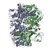 7ppeC 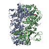 7ppfC 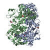 7pphC 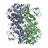 7ppiC 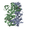 2gvjS S: Starting model for refinement C: citing same article ( |
|---|---|
| Similar structure data |
-Links
 PDBj
PDBj- Assembly
| Deposited unit | 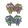
| ||||||||||||||||||||||||||||||||||||||||||||||||||||||||||||||||||||||||||||||||||||||||||||||||||
|---|---|---|---|---|---|---|---|---|---|---|---|---|---|---|---|---|---|---|---|---|---|---|---|---|---|---|---|---|---|---|---|---|---|---|---|---|---|---|---|---|---|---|---|---|---|---|---|---|---|---|---|---|---|---|---|---|---|---|---|---|---|---|---|---|---|---|---|---|---|---|---|---|---|---|---|---|---|---|---|---|---|---|---|---|---|---|---|---|---|---|---|---|---|---|---|---|---|---|---|
| 1 | 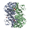
| ||||||||||||||||||||||||||||||||||||||||||||||||||||||||||||||||||||||||||||||||||||||||||||||||||
| 2 | 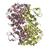
| ||||||||||||||||||||||||||||||||||||||||||||||||||||||||||||||||||||||||||||||||||||||||||||||||||
| Unit cell |
| ||||||||||||||||||||||||||||||||||||||||||||||||||||||||||||||||||||||||||||||||||||||||||||||||||
| Noncrystallographic symmetry (NCS) | NCS domain:
NCS domain segments: Component-ID: _ / Beg auth comp-ID: PHE / Beg label comp-ID: PHE / End auth comp-ID: ILE / End label comp-ID: ILE / Refine code: _ / Auth seq-ID: 9 - 484 / Label seq-ID: 10 - 485
NCS ensembles :
|
-Components
| #1: Protein | Mass: 55651.988 Da / Num. of mol.: 4 Source method: isolated from a genetically manipulated source Details: N-terminal Gly is a cloning artifact, stems from TEV cleavage site Source: (gene. exp.) Homo sapiens (human) / Gene: NAMPT, PBEF, PBEF1 / Production host:  References: UniProt: P43490, nicotinamide phosphoribosyltransferase #2: Chemical | ChemComp-PO4 /   Mass: 94.971 Da / Num. of mol.: 7 / Source method: obtained synthetically / Formula: PO4 Mass: 94.971 Da / Num. of mol.: 7 / Source method: obtained synthetically / Formula: PO4#3: Chemical | ChemComp-7YX / 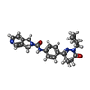  Mass: 417.503 Da / Num. of mol.: 4 / Source method: obtained synthetically / Formula: C24H27N5O2 / Feature type: SUBJECT OF INVESTIGATION Mass: 417.503 Da / Num. of mol.: 4 / Source method: obtained synthetically / Formula: C24H27N5O2 / Feature type: SUBJECT OF INVESTIGATION#4: Chemical | ChemComp-EDO /   Mass: 62.068 Da / Num. of mol.: 4 / Source method: obtained synthetically / Formula: C2H6O2 Mass: 62.068 Da / Num. of mol.: 4 / Source method: obtained synthetically / Formula: C2H6O2#5: Water | ChemComp-HOH / |  Mass: 18.015 Da / Num. of mol.: 1316 / Source method: isolated from a natural source / Formula: H2O Mass: 18.015 Da / Num. of mol.: 1316 / Source method: isolated from a natural source / Formula: H2OHas ligand of interest | Y | |
|---|
-Experimental details
-Experiment
| Experiment | Method: X-RAY DIFFRACTION / Number of used crystals: 1 |
|---|
- Sample preparation
| Crystal | Density Matthews: 2.39 Å3/Da / Density % sol: 48.43 % Description: Crescent-shaped, plate-shaped crystals, often with very uneven surfaces |
|---|---|
| Crystal grow | Temperature: 293 K / Method: vapor diffusion, hanging drop / pH: 7.6 Details: 1 microliter of protein mixed with 1 microliter of reservoir buffer (27-31% PEG 3350 (w/v), 200 mM NaCl, 100 mM sodium dihydrogen phosphate pH 7.6) incubated for 5 min, then streak seeded ...Details: 1 microliter of protein mixed with 1 microliter of reservoir buffer (27-31% PEG 3350 (w/v), 200 mM NaCl, 100 mM sodium dihydrogen phosphate pH 7.6) incubated for 5 min, then streak seeded (with crystals obtained previously under identical conditions). Ligand added prior to crystallization (2 MILLIMOLAR FROM 100 MILLIMOLAR STOCK IN DMSO) and incubated for 1.5 h at 277 K. CRYO BUFFER consisted of RESERVOIR supplemented WITH 2 MILLIMOLAR INHIBITOR AND 15% ETHYLENGLYCOL |
-Data collection
| Diffraction | Mean temperature: 100 K / Serial crystal experiment: N |
|---|---|
| Diffraction source | Source: SYNCHROTRON / Site: BESSY  / Beamline: 14.1 / Wavelength: 0.91841 Å / Beamline: 14.1 / Wavelength: 0.91841 Å |
| Detector | Type: DECTRIS PILATUS 6M / Detector: PIXEL / Date: Aug 28, 2014 |
| Radiation | Protocol: SINGLE WAVELENGTH / Monochromatic (M) / Laue (L): M / Scattering type: x-ray |
| Radiation wavelength | Wavelength: 0.91841 Å / Relative weight: 1 |
| Reflection | Resolution: 2.13→48.59 Å / Num. obs: 114934 / % possible obs: 98 % / Redundancy: 2.67 % / Biso Wilson estimate: 24.9 Å2 / CC1/2: 0.994 / Rrim(I) all: 0.151 / Rsym value: 0.121 / Net I/σ(I): 6.89 |
| Reflection shell | Resolution: 2.13→2.26 Å / Redundancy: 2.7 % / Mean I/σ(I) obs: 2.2 / Num. unique obs: 18131 / CC1/2: 0.854 / Rrim(I) all: 0.57 / Rsym value: 0.459 / % possible all: 96.1 |
- Processing
| Software |
| |||||||||||||||||||||||||||||||||||||||||||||||||||||||||||||||||
|---|---|---|---|---|---|---|---|---|---|---|---|---|---|---|---|---|---|---|---|---|---|---|---|---|---|---|---|---|---|---|---|---|---|---|---|---|---|---|---|---|---|---|---|---|---|---|---|---|---|---|---|---|---|---|---|---|---|---|---|---|---|---|---|---|---|---|
| Refinement | Method to determine structure: MOLECULAR REPLACEMENT Starting model: 2GVJ Resolution: 2.13→48.59 Å / Cor.coef. Fo:Fc: 0.934 / Cor.coef. Fo:Fc free: 0.897 / SU B: 8.165 / SU ML: 0.21 / SU R Cruickshank DPI: 0.3398 / Cross valid method: THROUGHOUT / σ(F): 0 / ESU R: 0.34 / ESU R Free: 0.256 / Stereochemistry target values: MAXIMUM LIKELIHOOD Details: HYDROGENS HAVE BEEN ADDED IN THE RIDING POSITIONS. U VALUES: REFINED INDIVIDUALLY.
| |||||||||||||||||||||||||||||||||||||||||||||||||||||||||||||||||
| Solvent computation | Ion probe radii: 0.8 Å / Shrinkage radii: 0.8 Å / VDW probe radii: 1.2 Å / Solvent model: MASK | |||||||||||||||||||||||||||||||||||||||||||||||||||||||||||||||||
| Displacement parameters | Biso max: 155.16 Å2 / Biso mean: 20.514 Å2 / Biso min: 0.5 Å2
| |||||||||||||||||||||||||||||||||||||||||||||||||||||||||||||||||
| Refinement step | Cycle: final / Resolution: 2.13→48.59 Å
| |||||||||||||||||||||||||||||||||||||||||||||||||||||||||||||||||
| Refine LS restraints |
| |||||||||||||||||||||||||||||||||||||||||||||||||||||||||||||||||
| Refine LS restraints NCS | Refine-ID: X-RAY DIFFRACTION / Type: interatomic distance / Weight position: 0.05
| |||||||||||||||||||||||||||||||||||||||||||||||||||||||||||||||||
| LS refinement shell | Resolution: 2.13→2.182 Å / Rfactor Rfree error: 0
|
