 Movie
Movie Controller

[English] 日本語
 Yorodumi
- PDB-7pnc: Dark state structure of Sensory Rhodopsin II solved by serial mil... -
+ Open data

- Basic information
| Entry | Database: PDB / ID: 7pnc | ||||||
|---|---|---|---|---|---|---|---|
| Title | Dark state structure of Sensory Rhodopsin II solved by serial millisecond crystallography | ||||||
 Components Components | Sensory rhodopsin-2 | ||||||
 Keywords Keywords | SIGNALING PROTEIN / SRII / serial millisecond crystallography / sensory rhodopsin / sensory rhodopsin II / SMX / SSX | ||||||
| Function / homology |  Function and homology information Function and homology informationphotoreceptor activity / phototransduction / monoatomic ion channel activity / plasma membrane Similarity search - Function | ||||||
| Biological species |  Natronomonas pharaonis (archaea) Natronomonas pharaonis (archaea) | ||||||
| Method |  X-RAY DIFFRACTION / SYNCHROTRON / MOLECULAR REPLACEMENT / Resolution: 2.2 Å X-RAY DIFFRACTION / SYNCHROTRON / MOLECULAR REPLACEMENT / Resolution: 2.2 Å | ||||||
 Authors Authors | Bosman, R. / Ortolani, G. / Branden, G. / Neutze, R. | ||||||
| Funding support | European Union, 1items
| ||||||
 Citation Citation | Journal: To Be Published Title: Structural basis of the prolonged photocycle of Sensory Rhodopsin II revealed by serial millisecond crystallography Authors: Bosman, R. / Ortolani, G. / Branden, G. / Neutze, R. | ||||||
| History |
|
- Structure visualization
| Structure viewer | Molecule: MolmilJmol/JSmol |
|---|
- Downloads & links
-Download
| PDBx/mmCIF format | 7pnc.cif.gz | 193.3 KB | Display | PDBx/mmCIF format |
|---|---|---|---|---|
| PDB format | pdb7pnc.ent.gz | 133.3 KB | Display | PDB format |
| PDBx/mmJSON format | 7pnc.json.gz | Tree view | PDBx/mmJSON format | |
| Others |  Other downloads Other downloads |
-Validation report
| Summary document | 7pnc_validation.pdf.gz | 2.7 MB | Display | wwPDB validaton report |
|---|---|---|---|---|
| Full document | 7pnc_full_validation.pdf.gz | 2.7 MB | Display | |
| Data in XML | 7pnc_validation.xml.gz | 13.3 KB | Display | |
| Data in CIF | 7pnc_validation.cif.gz | 16.9 KB | Display | |
| Arichive directory | https://data.pdbj.org/pub/pdb/validation_reports/pn/7pncftp://data.pdbj.org/pub/pdb/validation_reports/pn/7pnc | HTTPS FTP |
-Related structure data
| Related structure data |  8qqoC 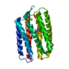 1h68S S: Starting model for refinement C: citing same article ( |
|---|---|
| Similar structure data |
-Links
 PDBj
PDBj


- Assembly
| Deposited unit | 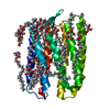
| ||||||||||||
|---|---|---|---|---|---|---|---|---|---|---|---|---|---|
| 1 |
| ||||||||||||
| Unit cell |
| ||||||||||||
| Components on special symmetry positions |
|
-Components
-Protein / Sugars , 2 types, 9 molecules A
| #1: Protein | Mass: 26666.137 Da / Num. of mol.: 1 Source method: isolated from a genetically manipulated source Source: (gene. exp.) Natronomonas pharaonis (archaea) / Gene: sop2, sopII / Production host:  |
|---|---|
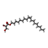
| #4: Sugar | ChemComp-BOG /  Type: D-saccharide / Mass: 292.369 Da / Num. of mol.: 8 / Source method: obtained synthetically / Formula: C14H28O6 / Comment: detergent*YM Type: D-saccharide / Mass: 292.369 Da / Num. of mol.: 8 / Source method: obtained synthetically / Formula: C14H28O6 / Comment: detergent*YM |
-Non-polymers , 4 types, 47 molecules 


| #2: Chemical | ChemComp-RET /  Mass: 284.436 Da / Num. of mol.: 1 / Source method: obtained synthetically / Formula: C20H28O / Feature type: SUBJECT OF INVESTIGATION Mass: 284.436 Da / Num. of mol.: 1 / Source method: obtained synthetically / Formula: C20H28O / Feature type: SUBJECT OF INVESTIGATION | ||||
|---|---|---|---|---|---|
| #3: Chemical |  Mass: 35.453 Da / Num. of mol.: 2 / Source method: obtained synthetically / Formula: Cl Mass: 35.453 Da / Num. of mol.: 2 / Source method: obtained synthetically / Formula: Cl#5: Chemical | ChemComp-MPG / [(  Mass: 356.540 Da / Num. of mol.: 6 / Source method: obtained synthetically / Formula: C21H40O4 Mass: 356.540 Da / Num. of mol.: 6 / Source method: obtained synthetically / Formula: C21H40O4#6: Water | ChemComp-HOH / | Mass: 18.015 Da / Num. of mol.: 38 / Source method: isolated from a natural source / Formula: H2O |
-Details
| Has ligand of interest | Y |
|---|
-Experimental details
-Experiment
| Experiment | Method: X-RAY DIFFRACTION / Number of used crystals: 1 |
|---|
- Sample preparation
| Crystal | Density Matthews: 2.8 Å3/Da / Density % sol: 56.04 % |
|---|---|
| Crystal grow | Temperature: 295 K / Method: lipidic cubic phase Details: CaCl 150mM, Glycine 100mM, 38%(v/v) PEG 400, pH 7.5 |
-Data collection
| Diffraction | Mean temperature: 298 K / Serial crystal experiment: Y |
|---|---|
| Diffraction source | Source: SYNCHROTRON / Site: SLS  / Beamline: X06SA / Wavelength: 1.0000342 Å / Beamline: X06SA / Wavelength: 1.0000342 Å |
| Detector | Type: DECTRIS EIGER X 16M / Detector: PIXEL / Date: May 15, 2019 |
| Radiation | Protocol: SINGLE WAVELENGTH / Monochromatic (M) / Laue (L): M / Scattering type: x-ray |
| Radiation wavelength | Wavelength: 1.0000342 Å / Relative weight: 1 |
| Reflection | Resolution: 2.1→39.44 Å / Num. obs: 18087 / % possible obs: 100 % / Redundancy: 242 % / Biso Wilson estimate: 33.33 Å2 / CC1/2: 0.991 / CC star: 0.998 / R split: 0.1399 / Net I/σ(I): 5.56 |
| Reflection shell | Resolution: 2.11→2.14 Å / Num. unique obs: 1722 / CC1/2: 0.31 / CC star: 0.686 |
| Serial crystallography sample delivery | Method: injection |
- Processing
| Software |
| |||||||||||||||||||||||||||||||||||||||||||||||||
|---|---|---|---|---|---|---|---|---|---|---|---|---|---|---|---|---|---|---|---|---|---|---|---|---|---|---|---|---|---|---|---|---|---|---|---|---|---|---|---|---|---|---|---|---|---|---|---|---|---|---|
| Refinement | Method to determine structure: MOLECULAR REPLACEMENT Starting model: 1H68 Resolution: 2.2→39.44 Å / SU ML: 0.1991 / Cross valid method: FREE R-VALUE / σ(F): 1.34 / Phase error: 21.5497 Stereochemistry target values: GeoStd + Monomer Library + CDL v1.2
| |||||||||||||||||||||||||||||||||||||||||||||||||
| Solvent computation | Shrinkage radii: 0.9 Å / VDW probe radii: 1.11 Å / Solvent model: FLAT BULK SOLVENT MODEL | |||||||||||||||||||||||||||||||||||||||||||||||||
| Displacement parameters | Biso mean: 55.66 Å2 | |||||||||||||||||||||||||||||||||||||||||||||||||
| Refinement step | Cycle: LAST / Resolution: 2.2→39.44 Å
| |||||||||||||||||||||||||||||||||||||||||||||||||
| Refine LS restraints |
| |||||||||||||||||||||||||||||||||||||||||||||||||
| LS refinement shell |
| |||||||||||||||||||||||||||||||||||||||||||||||||
| Refinement TLS params. | Method: refined / Origin x: 33.0000789233 Å / Origin y: 37.184319392 Å / Origin z: 2.1600188139 Å
| |||||||||||||||||||||||||||||||||||||||||||||||||
| Refinement TLS group | Selection details: all |
