 Movie
Movie Controller

[English] 日本語
 Yorodumi
- PDB-7pln: Structure of the APCbeta domain of Plasmodium vivax perforin-like... -
+ Open data

- Basic information
| Entry | Database: PDB / ID: 7pln | ||||||||||||
|---|---|---|---|---|---|---|---|---|---|---|---|---|---|
| Title | Structure of the APCbeta domain of Plasmodium vivax perforin-like protein 1 | ||||||||||||
 Components Components | Sporozoite micronemal protein essential for cell traversal, putative | ||||||||||||
 Keywords Keywords | CELL INVASION / Membrane binding domain / beta prism fold / APCbeta fold | ||||||||||||
| Function / homology | membrane-attack complex / perforin / MAC/Perforin domain / Membrane attack complex/perforin (MACPF) domain profile. / Membrane attack complex component/perforin (MACPF) domain / Sporozoite micronemal protein essential for cell traversal, putative Function and homology information Function and homology information | ||||||||||||
| Biological species |  | ||||||||||||
| Method |  X-RAY DIFFRACTION / SYNCHROTRON / MOLECULAR REPLACEMENT / Resolution: 3.15 Å X-RAY DIFFRACTION / SYNCHROTRON / MOLECULAR REPLACEMENT / Resolution: 3.15 Å | ||||||||||||
 Authors Authors | Williams, S.I. / Ni, T. / Yu, X. / Gilbert, R.J.C. | ||||||||||||
| Funding support |  United Kingdom, 3items United Kingdom, 3items
| ||||||||||||
 Citation Citation | Journal: J.Mol.Biol. / Year: 2022 Title: Structural, Functional and Computational Studies of Membrane Recognition by Plasmodium Perforin-Like Proteins 1 and 2 Authors: Williams, S.I. / Yu, X. / Ni, T. / Gilbert, R.J. / Stansfeld, P.J. | ||||||||||||
| History |
|
- Structure visualization
| Structure viewer | Molecule: MolmilJmol/JSmol |
|---|
- Downloads & links
-Download
| PDBx/mmCIF format | 7pln.cif.gz | 236.5 KB | Display | PDBx/mmCIF format |
|---|---|---|---|---|
| PDB format | pdb7pln.ent.gz | 160.8 KB | Display | PDB format |
| PDBx/mmJSON format | 7pln.json.gz | Tree view | PDBx/mmJSON format | |
| Others |  Other downloads Other downloads |
-Validation report
| Summary document | 7pln_validation.pdf.gz | 436.2 KB | Display | wwPDB validaton report |
|---|---|---|---|---|
| Full document | 7pln_full_validation.pdf.gz | 446.7 KB | Display | |
| Data in XML | 7pln_validation.xml.gz | 19.7 KB | Display | |
| Data in CIF | 7pln_validation.cif.gz | 26.2 KB | Display | |
| Arichive directory | https://data.pdbj.org/pub/pdb/validation_reports/pl/7plnftp://data.pdbj.org/pub/pdb/validation_reports/pl/7pln | HTTPS FTP |
-Related structure data
| Related structure data | 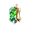 5ouoS S: Starting model for refinement |
|---|---|
| Similar structure data |
-Links
 PDBj
PDBj- Assembly
| Deposited unit | 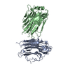
| ||||||||||||
|---|---|---|---|---|---|---|---|---|---|---|---|---|---|
| 1 | 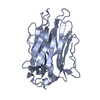
| ||||||||||||
| 2 | 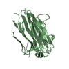
| ||||||||||||
| Unit cell |
|
-Components
| #1: Protein | Mass: 29784.506 Da / Num. of mol.: 2 Source method: isolated from a genetically manipulated source Details: PvPLP1 APCbeta domain polypeptide Source: (gene. exp.) Gene: PVC01_030012400, PVT01_030012800 / Cell line (production host): HEK293T / Production host:  Homo sapiens (human) / References: UniProt: A0A1G4H7E1 Homo sapiens (human) / References: UniProt: A0A1G4H7E1Has protein modification | Y | |
|---|
-Experimental details
-Experiment
| Experiment | Method: X-RAY DIFFRACTION / Number of used crystals: 1 |
|---|
- Sample preparation
| Crystal | Density Matthews: 2.18 Å3/Da / Density % sol: 43 % |
|---|---|
| Crystal grow | Temperature: 293 K / Method: vapor diffusion, sitting drop / Details: 20% PEG 3350, 0.2 M Tri-potassium citrate / Temp details: Room temperature |
-Data collection
| Diffraction | Mean temperature: 100 K / Serial crystal experiment: N |
|---|---|
| Diffraction source | Source: SYNCHROTRON / Site: Diamond / Beamline: I24 / Wavelength: 0.9686 Å |
| Detector | Type: DECTRIS PILATUS3 6M / Detector: PIXEL / Date: Jul 18, 2016 |
| Radiation | Protocol: SINGLE WAVELENGTH / Monochromatic (M) / Laue (L): M / Scattering type: x-ray |
| Radiation wavelength | Wavelength: 0.9686 Å / Relative weight: 1 |
| Reflection | Resolution: 3.15→63.01 Å / Num. obs: 9099 / % possible obs: 98.39 % / Observed criterion σ(F): 1 / Observed criterion σ(I): 1 / Redundancy: 13.5 % / Biso Wilson estimate: 85.93 Å2 / CC1/2: 0.995 / CC star: 0.999 / Rmerge(I) obs: 0.1942 / Rpim(I) all: 0.04997 / Rrim(I) all: 0.291 / Net I/σ(I): 11.32 |
| Reflection shell | Resolution: 3.15→3.263 Å / Redundancy: 2.8 % / Rmerge(I) obs: 0.5668 / Mean I/σ(I) obs: 1.68 / Num. unique obs: 790 / CC1/2: 0.695 / CC star: 0.906 / Rpim(I) all: 0.371 / % possible all: 84.67 |
- Processing
| Software |
| ||||||||||||||||||||||||||||||||||||||||
|---|---|---|---|---|---|---|---|---|---|---|---|---|---|---|---|---|---|---|---|---|---|---|---|---|---|---|---|---|---|---|---|---|---|---|---|---|---|---|---|---|---|
| Refinement | Method to determine structure: MOLECULAR REPLACEMENT Starting model: PDB 5OUO Resolution: 3.15→63.01 Å / SU ML: 0.4818 / Cross valid method: FREE R-VALUE / σ(F): 1.35 / Phase error: 32.1208 Stereochemistry target values: GeoStd + Monomer Library + CDL v1.2
| ||||||||||||||||||||||||||||||||||||||||
| Solvent computation | Shrinkage radii: 0.9 Å / VDW probe radii: 1.11 Å / Solvent model: FLAT BULK SOLVENT MODEL | ||||||||||||||||||||||||||||||||||||||||
| Displacement parameters | Biso mean: 84.11 Å2 | ||||||||||||||||||||||||||||||||||||||||
| Refinement step | Cycle: LAST / Resolution: 3.15→63.01 Å
| ||||||||||||||||||||||||||||||||||||||||
| Refine LS restraints |
| ||||||||||||||||||||||||||||||||||||||||
| LS refinement shell |
| ||||||||||||||||||||||||||||||||||||||||
| Refinement TLS params. | Method: refined / Origin x: 0.900807212513 Å / Origin y: 14.6180109927 Å / Origin z: -22.3290329102 Å
| ||||||||||||||||||||||||||||||||||||||||
| Refinement TLS group | Selection details: all |
