 Movie
Movie Controller


+ Open data

- Basic information
| Entry | Database: PDB / ID: 7pfh | ||||||
|---|---|---|---|---|---|---|---|
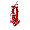
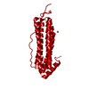
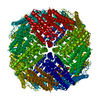
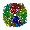
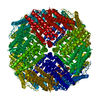
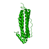
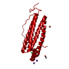
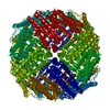
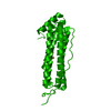
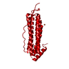
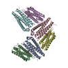
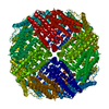
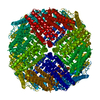
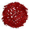
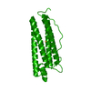
| Title | 2 minute Fe2+ soak structure of SynFtn E141D | ||||||
 Components Components | Ferritin | ||||||
 Keywords Keywords | METAL BINDING PROTEIN / iron binding protein | ||||||
| Function / homology |  Function and homology information Function and homology informationbacterial non-heme ferritin / ferric iron binding / iron ion transport / ferrous iron binding / intracellular iron ion homeostasis / cytoplasm Similarity search - Function | ||||||
| Biological species |  Synechococcus sp. (bacteria) Synechococcus sp. (bacteria) | ||||||
| Method |  X-RAY DIFFRACTION / SYNCHROTRON / MOLECULAR REPLACEMENT / Resolution: 1.5 Å X-RAY DIFFRACTION / SYNCHROTRON / MOLECULAR REPLACEMENT / Resolution: 1.5 Å | ||||||
| Model details | structure determined from crystals of SynFtn D65A that have not been soaked in Fe2+ | ||||||
 Authors Authors | Hemmings, A.M. / Bradley, J.M. | ||||||
| Funding support |  United Kingdom, 1items United Kingdom, 1items
| ||||||
 Citation Citation | Journal: Microbiology (Reading, Engl.) / Year: 2021 Title: Key carboxylate residues for iron transit through the prokaryotic ferritin Syn Ftn. Authors: Bradley, J.M. / Fair, J. / Hemmings, A.M. / Le Brun, N.E. | ||||||
| History |
|
- Structure visualization
| Structure viewer | Molecule: MolmilJmol/JSmol |
|---|
- Downloads & links
-Download
| PDBx/mmCIF format | 7pfh.cif.gz | 90.6 KB | Display | PDBx/mmCIF format |
|---|---|---|---|---|
| PDB format | pdb7pfh.ent.gz | 69.1 KB | Display | PDB format |
| PDBx/mmJSON format | 7pfh.json.gz | Tree view | PDBx/mmJSON format | |
| Others |  Other downloads Other downloads |
-Validation report
| Arichive directory | https://data.pdbj.org/pub/pdb/validation_reports/pf/7pfhftp://data.pdbj.org/pub/pdb/validation_reports/pf/7pfh | HTTPS FTP |
|---|
-Related structure data
| Related structure data | 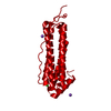 7pf7C 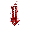 7pf8C 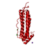 7pf9C  7pfbC 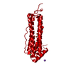 7pfgC  7pfiC  7pfjC  7pfkC 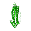 5ouwS C: citing same article ( S: Starting model for refinement |
|---|---|
| Similar structure data |


-Links
 PDBj
PDBj


- Assembly
| Deposited unit | 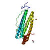
| ||||||||||||||||||
|---|---|---|---|---|---|---|---|---|---|---|---|---|---|---|---|---|---|---|---|
| 1 | 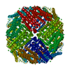
| ||||||||||||||||||
| Unit cell |
| ||||||||||||||||||
| Components on special symmetry positions |
|
-Components
| #1: Protein | Mass: 20218.330 Da / Num. of mol.: 1 / Fragment: ferritin / Mutation: E141D Source method: isolated from a genetically manipulated source Source: (gene. exp.) Synechococcus sp. (strain CC9311) (bacteria)Strain: CC9311 / Gene: sync_1539 / Plasmid: pET21a / Production host: | ||||||||
|---|---|---|---|---|---|---|---|---|---|
| #2: Chemical |   Mass: 55.845 Da / Num. of mol.: 3 / Source method: obtained synthetically / Formula: Fe / Feature type: SUBJECT OF INVESTIGATION Mass: 55.845 Da / Num. of mol.: 3 / Source method: obtained synthetically / Formula: Fe / Feature type: SUBJECT OF INVESTIGATION#3: Chemical |   Mass: 35.453 Da / Num. of mol.: 3 / Source method: obtained synthetically / Formula: Cl Mass: 35.453 Da / Num. of mol.: 3 / Source method: obtained synthetically / Formula: Cl#4: Chemical | ChemComp-ACT / |   Mass: 59.044 Da / Num. of mol.: 1 / Source method: obtained synthetically / Formula: C2H3O2 Mass: 59.044 Da / Num. of mol.: 1 / Source method: obtained synthetically / Formula: C2H3O2#5: Water | ChemComp-HOH / |  Mass: 18.015 Da / Num. of mol.: 203 / Source method: isolated from a natural source / Formula: H2O Mass: 18.015 Da / Num. of mol.: 203 / Source method: isolated from a natural source / Formula: H2OHas ligand of interest | Y | |
-Experimental details
-Experiment
| Experiment | Method: X-RAY DIFFRACTION / Number of used crystals: 1 |
|---|
- Sample preparation
| Crystal | Density Matthews: 2.85 Å3/Da / Density % sol: 56.85 % / Mosaicity: 0.051 ° |
|---|---|
| Crystal grow | Temperature: 289 K / Method: vapor diffusion, sitting drop / pH: 4.6 / Details: 2M NaCl |
-Data collection
| Diffraction | Mean temperature: 100 K / Serial crystal experiment: N | ||||||||||||||||||||||||
|---|---|---|---|---|---|---|---|---|---|---|---|---|---|---|---|---|---|---|---|---|---|---|---|---|---|
| Diffraction source | Source: SYNCHROTRON / Site: Diamond / Beamline: I03 / Wavelength: 0.9763 Å | ||||||||||||||||||||||||
| Detector | Type: DECTRIS EIGER X 16M / Detector: PIXEL / Date: Jun 17, 2020 | ||||||||||||||||||||||||
| Radiation | Protocol: SINGLE WAVELENGTH / Monochromatic (M) / Laue (L): M / Scattering type: x-ray | ||||||||||||||||||||||||
| Radiation wavelength | Wavelength: 0.9763 Å / Relative weight: 1 | ||||||||||||||||||||||||
| Reflection | Resolution: 1.41→53.3 Å / Num. obs: 45787 / % possible obs: 99.7 % / Redundancy: 15 % / Rpim(I) all: 0.011 / Rrim(I) all: 0.047 / Net I/σ(I): 23.3 / Num. measured all: 685729 | ||||||||||||||||||||||||
| Reflection shell | Diffraction-ID: 1
|
- Processing
| Software |
| ||||||||||||||||||||||||||||||||||||||||||||||||||||||||||||||||||||||||||||||||||||
|---|---|---|---|---|---|---|---|---|---|---|---|---|---|---|---|---|---|---|---|---|---|---|---|---|---|---|---|---|---|---|---|---|---|---|---|---|---|---|---|---|---|---|---|---|---|---|---|---|---|---|---|---|---|---|---|---|---|---|---|---|---|---|---|---|---|---|---|---|---|---|---|---|---|---|---|---|---|---|---|---|---|---|---|---|---|
| Refinement | Method to determine structure: MOLECULAR REPLACEMENT Starting model: 5OUW Resolution: 1.5→44.218 Å / SU ML: 0.12 / Cross valid method: THROUGHOUT / σ(F): 1.34 / Phase error: 15.97 / Stereochemistry target values: ML
| ||||||||||||||||||||||||||||||||||||||||||||||||||||||||||||||||||||||||||||||||||||
| Solvent computation | Shrinkage radii: 0.9 Å / VDW probe radii: 1.11 Å / Solvent model: FLAT BULK SOLVENT MODEL | ||||||||||||||||||||||||||||||||||||||||||||||||||||||||||||||||||||||||||||||||||||
| Displacement parameters | Biso max: 73.7 Å2 / Biso mean: 30.7856 Å2 / Biso min: 21.6 Å2 | ||||||||||||||||||||||||||||||||||||||||||||||||||||||||||||||||||||||||||||||||||||
| Refinement step | Cycle: final / Resolution: 1.5→44.218 Å
| ||||||||||||||||||||||||||||||||||||||||||||||||||||||||||||||||||||||||||||||||||||
| Refine LS restraints |
| ||||||||||||||||||||||||||||||||||||||||||||||||||||||||||||||||||||||||||||||||||||
| LS refinement shell | Refine-ID: X-RAY DIFFRACTION / Rfactor Rfree error: 0
|
