 Movie
Movie Controller

[English] 日本語
 Yorodumi
- PDB-7pce: BurG (apo): Biosynthesis of cyclopropanol rings in bacterial toxins -
+ Open data

- Basic information
| Entry | Database: PDB / ID: 7pce | ||||||
|---|---|---|---|---|---|---|---|
| Title | BurG (apo): Biosynthesis of cyclopropanol rings in bacterial toxins | ||||||
 Components Components | Ketol-acid reductoisomerase | ||||||
 Keywords Keywords | LYASE / Pathogens / Natural Products / Toxins / Biosynthesis / Catalysis | ||||||
| Function / homology |  Function and homology information Function and homology informationketol-acid reductoisomerase (NADP+) / ketol-acid reductoisomerase activity / L-valine biosynthetic process / : / nucleotide binding / metal ion binding Similarity search - Function | ||||||
| Biological species |  Burkholderia thailandensis (bacteria) Burkholderia thailandensis (bacteria) | ||||||
| Method |  X-RAY DIFFRACTION / SYNCHROTRON / MOLECULAR REPLACEMENT / Resolution: 2.9 Å X-RAY DIFFRACTION / SYNCHROTRON / MOLECULAR REPLACEMENT / Resolution: 2.9 Å | ||||||
 Authors Authors | Trottmann, F. / Ishida, K. / Ishida, M. / Kries, H. / Groll, M. / Hertweck, C. | ||||||
| Funding support |  Germany, 1items Germany, 1items
| ||||||
 Citation Citation | Journal: Nat.Chem. / Year: 2022 Title: Pathogenic bacteria remodel central metabolic enzyme to build a cyclopropanol warhead. Authors: Trottmann, F. / Ishida, K. / Ishida-Ito, M. / Kries, H. / Groll, M. / Hertweck, C. | ||||||
| History |
|
- Structure visualization
| Structure viewer | Molecule: MolmilJmol/JSmol |
|---|
- Downloads & links
-Download
| PDBx/mmCIF format | 7pce.cif.gz | 138.8 KB | Display | PDBx/mmCIF format |
|---|---|---|---|---|
| PDB format | pdb7pce.ent.gz | 107.9 KB | Display | PDB format |
| PDBx/mmJSON format | 7pce.json.gz | Tree view | PDBx/mmJSON format | |
| Others |  Other downloads Other downloads |
-Validation report
| Arichive directory | https://data.pdbj.org/pub/pdb/validation_reports/pc/7pceftp://data.pdbj.org/pub/pdb/validation_reports/pc/7pce | HTTPS FTP |
|---|
-Related structure data
| Related structure data | 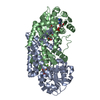 7pccSC 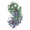 7pcgC 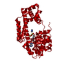 7pciC  7pclC 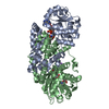 7pcmC 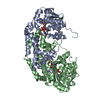 7pcnC 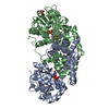 7pcoC  7pctC S: Starting model for refinement C: citing same article ( |
|---|---|
| Similar structure data |

-Links
 PDBj
PDBj


- Assembly
| Deposited unit | 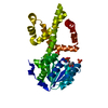
| ||||||||
|---|---|---|---|---|---|---|---|---|---|
| 1 | 
| ||||||||
| Unit cell |
|
-Components
| #1: Protein | Mass: 38716.699 Da / Num. of mol.: 1 Source method: isolated from a genetically manipulated source Source: (gene. exp.) Burkholderia thailandensis (strain ATCC 700388 / DSM 13276 / CIP 106301 / E264) (bacteria)Strain: ATCC 700388 / DSM 13276 / CIP 106301 / E264 / Gene: ilvC-2, BTH_II2094 / Production host: References: UniProt: Q2T3G7, ketol-acid reductoisomerase (NADP+) | ||
|---|---|---|---|
| #2: Chemical | ChemComp-ADP /   Mass: 427.201 Da / Num. of mol.: 1 / Source method: obtained synthetically / Formula: C10H15N5O10P2 / Comment: ADP, energy-carrying molecule*YM Mass: 427.201 Da / Num. of mol.: 1 / Source method: obtained synthetically / Formula: C10H15N5O10P2 / Comment: ADP, energy-carrying molecule*YM | ||
| #3: Chemical |   Mass: 94.971 Da / Num. of mol.: 2 / Source method: obtained synthetically / Formula: PO4 Mass: 94.971 Da / Num. of mol.: 2 / Source method: obtained synthetically / Formula: PO4Has ligand of interest | N | |
-Experimental details
-Experiment
| Experiment | Method: X-RAY DIFFRACTION / Number of used crystals: 1 |
|---|
- Sample preparation
| Crystal | Density Matthews: 2.67 Å3/Da / Density % sol: 53.86 % |
|---|---|
| Crystal grow | Temperature: 293 K / Method: vapor diffusion, sitting drop / pH: 6.2 / Details: 0.1 M Na/K-phosphate; 0.2 M NaCl; 50% PEG 200 |
-Data collection
| Diffraction | Mean temperature: 100 K / Serial crystal experiment: N |
|---|---|
| Diffraction source | Source: SYNCHROTRON / Site: SLS  / Beamline: X06SA / Wavelength: 1 Å / Beamline: X06SA / Wavelength: 1 Å |
| Detector | Type: DECTRIS EIGER2 X 16M / Detector: PIXEL / Date: Dec 14, 2020 |
| Radiation | Protocol: SINGLE WAVELENGTH / Monochromatic (M) / Laue (L): M / Scattering type: x-ray |
| Radiation wavelength | Wavelength: 1 Å / Relative weight: 1 |
| Reflection | Resolution: 2.9→30 Å / Num. obs: 9678 / % possible obs: 99 % / Redundancy: 4.8 % / Rmerge(I) obs: 0.065 / Net I/σ(I): 14.2 |
| Reflection shell | Resolution: 2.9→3 Å / Rmerge(I) obs: 0.588 / Mean I/σ(I) obs: 2 / Num. unique obs: 884 |
- Processing
| Software |
| ||||||||||||||||||||||||||||||||||||||||||||||||||||||||||||
|---|---|---|---|---|---|---|---|---|---|---|---|---|---|---|---|---|---|---|---|---|---|---|---|---|---|---|---|---|---|---|---|---|---|---|---|---|---|---|---|---|---|---|---|---|---|---|---|---|---|---|---|---|---|---|---|---|---|---|---|---|---|
| Refinement | Method to determine structure: MOLECULAR REPLACEMENT Starting model: 7PCC Resolution: 2.9→30 Å / Cor.coef. Fo:Fc: 0.948 / Cor.coef. Fo:Fc free: 0.939 / SU B: 35.875 / SU ML: 0.286 / Cross valid method: THROUGHOUT / σ(F): 0 / ESU R Free: 0.383 / Stereochemistry target values: MAXIMUM LIKELIHOOD Details: HYDROGENS HAVE BEEN ADDED IN THE RIDING POSITIONS U VALUES : WITH TLS ADDED
| ||||||||||||||||||||||||||||||||||||||||||||||||||||||||||||
| Solvent computation | Ion probe radii: 0.8 Å / Shrinkage radii: 0.8 Å / VDW probe radii: 1.2 Å / Solvent model: MASK | ||||||||||||||||||||||||||||||||||||||||||||||||||||||||||||
| Displacement parameters | Biso max: 130.5 Å2 / Biso mean: 79.874 Å2 / Biso min: 69.23 Å2
| ||||||||||||||||||||||||||||||||||||||||||||||||||||||||||||
| Refinement step | Cycle: final / Resolution: 2.9→30 Å
| ||||||||||||||||||||||||||||||||||||||||||||||||||||||||||||
| Refine LS restraints |
| ||||||||||||||||||||||||||||||||||||||||||||||||||||||||||||
| LS refinement shell | Resolution: 2.9→2.975 Å / Rfactor Rfree error: 0 / Total num. of bins used: 20
| ||||||||||||||||||||||||||||||||||||||||||||||||||||||||||||
| Refinement TLS params. | Method: refined / Origin x: -6.8509 Å / Origin y: 17.3794 Å / Origin z: 16.5367 Å
|
