- PDB-7p8v: The structure of E. coli MutL bound to a 3' resected DNA end -
+
Open data
ID or keywords:
Loading...
-
Basic information
Entry
Database: PDB / ID: 7p8v
Title
The structure of E. coli MutL bound to a 3' resected DNA end
Components
DNA mismatch repair protein MutL
Primer strand
Template strand
Keywords
DNA BINDING PROTEIN / DNA mismatch repair / Protein-DNA complex
Function / homology
Function and homology information
single-stranded DNA-dependent ATP-dependent DNA helicase complex / mismatch repair involved in maintenance of fidelity involved in DNA-dependent DNA replication / mismatch repair complex / regulation of DNA recombination / mismatched DNA binding / ATP-dependent DNA damage sensor activity / mismatch repair / ATP hydrolysis activity / DNA binding / ATP binding / identical protein binding Similarity search - Function
DNA mismatch repair protein, MutL / MutL, C-terminal domain, regulatory subdomain / MutL C terminal dimerisation domain / MutL, C-terminal, dimerisation / MutL, C-terminal domain superfamily / MutL, C-terminal domain, dimerisation subdomain / MutL C terminal dimerisation domain / DNA mismatch repair protein family, N-terminal / DNA mismatch repair protein, S5 domain 2-like / DNA mismatch repair, conserved site ...DNA mismatch repair protein, MutL / MutL, C-terminal domain, regulatory subdomain / MutL C terminal dimerisation domain / MutL, C-terminal, dimerisation / MutL, C-terminal domain superfamily / MutL, C-terminal domain, dimerisation subdomain / MutL C terminal dimerisation domain / DNA mismatch repair protein family, N-terminal / DNA mismatch repair protein, S5 domain 2-like / DNA mismatch repair, conserved site / DNA mismatch repair protein MutL/Mlh/Pms / DNA mismatch repair protein, C-terminal domain / DNA mismatch repair proteins mutL / hexB / PMS1 signature. / DNA mismatch repair protein, C-terminal domain / Histidine kinase-, DNA gyrase B-, and HSP90-like ATPase / Histidine kinase/HSP90-like ATPase superfamily / Ribosomal protein S5 domain 2-type fold, subgroup / Ribosomal protein S5 domain 2-type fold Similarity search - Domain/homology
PHOSPHOAMINOPHOSPHONIC ACID-ADENYLATE ESTER / DNA / DNA (> 10) / DNA mismatch repair protein MutL Similarity search - Component
Biological species
Escherichia coli (E. coli) DNA molecule (others)
Method
ELECTRON MICROSCOPY / single particle reconstruction / cryo EM / Resolution: 3.6 Å
Journal: Nucleic Acids Res / Year: 2022 Title: MutL binds to 3' resected DNA ends and blocks DNA polymerase access. Authors: Alessandro Borsellini / Joyce H G Lebbink / Meindert H Lamers / Abstract: DNA mismatch repair removes mis-incorporated bases after DNA replication and reduces the error rate a 100-1000-fold. After recognition of a mismatch, a large section of up to a thousand nucleotides ...DNA mismatch repair removes mis-incorporated bases after DNA replication and reduces the error rate a 100-1000-fold. After recognition of a mismatch, a large section of up to a thousand nucleotides is removed from the daughter strand followed by re-synthesis. How these opposite activities are coordinated is poorly understood. Here we show that the Escherichia coli MutL protein binds to the 3' end of the resected strand and blocks access of Pol I and Pol III. The cryo-EM structure of an 85-kDa MutL-DNA complex, determined to 3.7 Å resolution, reveals a unique DNA binding mode that positions MutL at the 3' end of a primer-template, but not at a 5' resected DNA end or a blunt DNA end. Hence, our work reveals a novel role for MutL in the final stages of mismatch repair by preventing premature DNA synthesis during removal of the mismatched strand.
In the structure databanks used in Yorodumi, some data are registered as the other names, "COVID-19 virus" and "2019-nCoV". Here are the details of the virus and the list of structure data.
Jan 31, 2019. EMDB accession codes are about to change! (news from PDBe EMDB page)
EMDB accession codes are about to change! (news from PDBe EMDB page)
The allocation of 4 digits for EMDB accession codes will soon come to an end. Whilst these codes will remain in use, new EMDB accession codes will include an additional digit and will expand incrementally as the available range of codes is exhausted. The current 4-digit format prefixed with “EMD-” (i.e. EMD-XXXX) will advance to a 5-digit format (i.e. EMD-XXXXX), and so on. It is currently estimated that the 4-digit codes will be depleted around Spring 2019, at which point the 5-digit format will come into force.
The EM Navigator/Yorodumi systems omit the EMD- prefix.
Related info.:Q: What is EMD? / ID/Accession-code notation in Yorodumi/EM Navigator
Yorodumi is a browser for structure data from EMDB, PDB, SASBDB, etc.
This page is also the successor to EM Navigator detail page, and also detail information page/front-end page for Omokage search.
The word "yorodu" (or yorozu) is an old Japanese word meaning "ten thousand". "mi" (miru) is to see.
Related info.:EMDB / PDB / SASBDB / Comparison of 3 databanks / Yorodumi Search / Aug 31, 2016. New EM Navigator & Yorodumi / Yorodumi Papers / Jmol/JSmol / Function and homology information / Changes in new EM Navigator and Yorodumi
 Movie
Movie Controller


 Open data
 Basic information
 Components
 Keywords
 Function and homology information
 Authors
 Citation
 Structure visualization
 Downloads & links
 Other downloads
 PDBj
PDBj


 Assembly
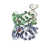

 Mass: 506.196 Da / Num. of mol.: 2 / Source method: obtained synthetically / Formula: C10H17N6O12P3 / Comment: AMP-PNP, energy-carrying molecule analogue*YM
 Mass: 24.305 Da / Num. of mol.: 2 / Source method: obtained synthetically / Formula: Mg
 Sample preparation
 Electron microscopy imaging
 FIELD EMISSION GUN / Accelerating voltage: 300 kV / Illumination mode: FLOOD BEAM
 Processing