 Movie
Movie Controller

[English] 日本語
 Yorodumi
- PDB-7p8j: Receptor-binding domain (RBD) of the spike protein of the bat cor... -
+ Open data

- Basic information
| Entry | Database: PDB / ID: 7p8j | ||||||||||||||||||||||||
|---|---|---|---|---|---|---|---|---|---|---|---|---|---|---|---|---|---|---|---|---|---|---|---|---|---|
| Title | Receptor-binding domain (RBD) of the spike protein of the bat coronavirus RaTG13 virus in complex with the extracellular domain of human angiotensin-converting enzyme 2 (ACE2) - Crystal form 2 | ||||||||||||||||||||||||
 Components Components |
| ||||||||||||||||||||||||
 Keywords Keywords | CELL INVASION / COVID-19 / Bat coronavirus / Spillover / Zoonotic viral infection / Evolution | ||||||||||||||||||||||||
| Function / homology |  Function and homology information Function and homology informationpositive regulation of amino acid transport / angiotensin-converting enzyme 2 / positive regulation of L-proline import across plasma membrane / Hydrolases; Acting on peptide bonds (peptidases); Metallocarboxypeptidases / angiotensin-mediated drinking behavior / positive regulation of gap junction assembly / regulation of systemic arterial blood pressure by renin-angiotensin / tryptophan transport / regulation of cardiac conduction / maternal process involved in female pregnancy ...positive regulation of amino acid transport / angiotensin-converting enzyme 2 / positive regulation of L-proline import across plasma membrane / Hydrolases; Acting on peptide bonds (peptidases); Metallocarboxypeptidases / angiotensin-mediated drinking behavior / positive regulation of gap junction assembly / regulation of systemic arterial blood pressure by renin-angiotensin / tryptophan transport / regulation of cardiac conduction / maternal process involved in female pregnancy / peptidyl-dipeptidase activity / regulation of vasoconstriction / transporter activator activity / Metabolism of Angiotensinogen to Angiotensins / carboxypeptidase activity / angiotensin maturation / viral life cycle / Attachment and Entry / receptor-mediated endocytosis of virus by host cell / metallocarboxypeptidase activity / positive regulation of cardiac muscle contraction / regulation of cytokine production / blood vessel diameter maintenance / negative regulation of smooth muscle cell proliferation / brush border membrane / negative regulation of ERK1 and ERK2 cascade / positive regulation of reactive oxygen species metabolic process / metallopeptidase activity / endocytic vesicle membrane / regulation of cell population proliferation / virus receptor activity / regulation of inflammatory response / endopeptidase activity / viral translation / Potential therapeutics for SARS / Induction of Cell-Cell Fusion / membrane fusion / entry receptor-mediated virion attachment to host cell / Attachment and Entry / host cell endoplasmic reticulum-Golgi intermediate compartment membrane / receptor-mediated virion attachment to host cell / cilium / apical plasma membrane / membrane raft / endocytosis involved in viral entry into host cell / endoplasmic reticulum lumen / fusion of virus membrane with host plasma membrane / fusion of virus membrane with host endosome membrane / viral envelope / symbiont entry into host cell / host cell plasma membrane / virion membrane / cell surface / negative regulation of transcription by RNA polymerase II / extracellular space / extracellular exosome / extracellular region / zinc ion binding / identical protein binding / membrane / plasma membrane Similarity search - Function | ||||||||||||||||||||||||
| Biological species |  Homo sapiens (human) Homo sapiens (human) Bat coronavirus RaTG13 Bat coronavirus RaTG13 | ||||||||||||||||||||||||
| Method |  X-RAY DIFFRACTION / SYNCHROTRON / MOLECULAR REPLACEMENT / Resolution: 6.585 Å X-RAY DIFFRACTION / SYNCHROTRON / MOLECULAR REPLACEMENT / Resolution: 6.585 Å | ||||||||||||||||||||||||
 Authors Authors | Scietti, L. / Castelli, M. / Faravelli, S. / Clementi, N. / Mancini, N. / Forneris, F. | ||||||||||||||||||||||||
| Funding support |  Italy, Italy,  United States, United States,  Switzerland, Switzerland,  Japan, Japan,  Belgium, 7items Belgium, 7items
| ||||||||||||||||||||||||
 Citation Citation | Journal: To Be Published Title: Constrained Evolution of SARS-CoV-2 Spike in Rhinolophus affinis Bats Authors: Castelli, M. / Scietti, L. / Faravelli, S. / Clementi, N. / Forneris, F. / Mancini, N. | ||||||||||||||||||||||||
| History |
|
- Structure visualization
| Structure viewer | Molecule: MolmilJmol/JSmol |
|---|
- Downloads & links
-Download
| PDBx/mmCIF format | 7p8j.cif.gz | 298.7 KB | Display | PDBx/mmCIF format |
|---|---|---|---|---|
| PDB format | pdb7p8j.ent.gz | 228.7 KB | Display | PDB format |
| PDBx/mmJSON format | 7p8j.json.gz | Tree view | PDBx/mmJSON format | |
| Others |  Other downloads Other downloads |
-Validation report
| Arichive directory | https://data.pdbj.org/pub/pdb/validation_reports/p8/7p8jftp://data.pdbj.org/pub/pdb/validation_reports/p8/7p8j | HTTPS FTP |
|---|
-Related structure data
| Related structure data |  6vw1S S: Starting model for refinement |
|---|---|
| Similar structure data |
-Links
 PDBj
PDBj


- Assembly
| Deposited unit | 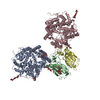
| ||||||||||||||||||||||||||||||||||||||||||||||||
|---|---|---|---|---|---|---|---|---|---|---|---|---|---|---|---|---|---|---|---|---|---|---|---|---|---|---|---|---|---|---|---|---|---|---|---|---|---|---|---|---|---|---|---|---|---|---|---|---|---|
| 1 | 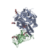
| ||||||||||||||||||||||||||||||||||||||||||||||||
| 2 | 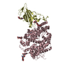
| ||||||||||||||||||||||||||||||||||||||||||||||||
| Unit cell |
| ||||||||||||||||||||||||||||||||||||||||||||||||
| Noncrystallographic symmetry (NCS) | NCS domain:
NCS domain segments:
NCS ensembles :
|
-Components
| #1: Protein | Mass: 69511.039 Da / Num. of mol.: 2 Source method: isolated from a genetically manipulated source Details: Initial GS and final AAA sequences were introduced by molecular cloning Source: (gene. exp.) Homo sapiens (human) / Gene: ACE2, UNQ868/PRO1885 / Plasmid: pUPE.06.45 / Details (production host): N-term His-Strep-TEV / Cell line (production host): HEK293F / Production host: Homo sapiens (human) / References: UniProt: Q9BYF1#2: Protein | Mass: 26265.598 Da / Num. of mol.: 2 Source method: isolated from a genetically manipulated source Details: cloning artifacts: GS sequence at the N-terminus and AA sequence at the C-terminus, + 6xHis-tag at the C-terminus Source: (gene. exp.) Bat coronavirus RaTG13 / Plasmid: pCAGGS / Cell line (production host): HEK293F / Production host: Homo sapiens (human) / References: UniProt: A0A6B9WHD3#3: Polysaccharide | 2-acetamido-2-deoxy-beta-D-glucopyranose-(1-4)-2-acetamido-2-deoxy-beta-D-glucopyranose Source method: isolated from a genetically manipulated source #4: Polysaccharide | beta-D-mannopyranose-(1-4)-2-acetamido-2-deoxy-beta-D-glucopyranose-(1-4)-2-acetamido-2-deoxy-beta- ...beta-D-mannopyranose-(1-4)-2-acetamido-2-deoxy-beta-D-glucopyranose-(1-4)-2-acetamido-2-deoxy-beta-D-glucopyranose Source method: isolated from a genetically manipulated source Has ligand of interest | N | Has protein modification | Y | |
|---|
-Experimental details
-Experiment
| Experiment | Method: X-RAY DIFFRACTION / Number of used crystals: 1 |
|---|
- Sample preparation
| Crystal | Density Matthews: 3.18 Å3/Da / Density % sol: 61.34 % |
|---|---|
| Crystal grow | Temperature: 293 K / Method: vapor diffusion / pH: 6.9 Details: 0.2-0.25 M sodium thiocyanate, 18-23% PEG 3350, pH 6.9 |
-Data collection
| Diffraction | Mean temperature: 100 K / Serial crystal experiment: N |
|---|---|
| Diffraction source | Source: SYNCHROTRON / Site: ESRF  / Beamline: ID23-1 / Wavelength: 1 Å / Beamline: ID23-1 / Wavelength: 1 Å |
| Detector | Type: DECTRIS PILATUS 6M / Detector: PIXEL / Date: Feb 13, 2021 |
| Radiation | Protocol: SINGLE WAVELENGTH / Monochromatic (M) / Laue (L): M / Scattering type: x-ray |
| Radiation wavelength | Wavelength: 1 Å / Relative weight: 1 |
| Reflection | Resolution: 6.5→66.42 Å / Num. obs: 5280 / % possible obs: 99.7 % / Redundancy: 8.5 % / CC1/2: 0.979 / Rmerge(I) obs: 0.459 / Net I/σ(I): 2.8 |
| Reflection shell | Resolution: 6.5→7.26 Å / Redundancy: 8.9 % / Rmerge(I) obs: 2.905 / Mean I/σ(I) obs: 0.4 / Num. unique obs: 1424 / CC1/2: 0.531 / % possible all: 99.2 |
- Processing
| Software |
| |||||||||||||||||||||||||||||||||||
|---|---|---|---|---|---|---|---|---|---|---|---|---|---|---|---|---|---|---|---|---|---|---|---|---|---|---|---|---|---|---|---|---|---|---|---|---|
| Refinement | Method to determine structure: MOLECULAR REPLACEMENT Starting model: 6VW1 Resolution: 6.585→49.516 Å / SU ML: 0.89 / Cross valid method: THROUGHOUT / σ(F): 1.34 / Phase error: 34.86 / Stereochemistry target values: ML
| |||||||||||||||||||||||||||||||||||
| Solvent computation | Shrinkage radii: 0.9 Å / VDW probe radii: 1.11 Å / Solvent model: FLAT BULK SOLVENT MODEL | |||||||||||||||||||||||||||||||||||
| Displacement parameters | Biso max: 30 Å2 / Biso mean: 30 Å2 / Biso min: 30 Å2 | |||||||||||||||||||||||||||||||||||
| Refinement step | Cycle: final / Resolution: 6.585→49.516 Å
| |||||||||||||||||||||||||||||||||||
| Refine LS restraints NCS |
| |||||||||||||||||||||||||||||||||||
| LS refinement shell | Resolution: 6.5855→49.516 Å / Rfactor Rfree error: 0
|
