 Movie
Movie Controller

[English] 日本語
 Yorodumi
- PDB-7p4w: Crystal structure of alpha-amylase from Aspergillus oryzae in spa... -
+ Open data

- Basic information
| Entry | Database: PDB / ID: 7p4w | ||||||
|---|---|---|---|---|---|---|---|
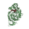
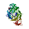
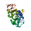
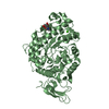
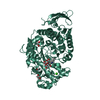
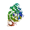
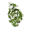
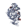
| Title | Crystal structure of alpha-amylase from Aspergillus oryzae in space group I222 | ||||||
 Components Components | Alpha-amylase | ||||||
 Keywords Keywords | HYDROLASE / amylase / aspergillus / oryzae / alpha-amylase | ||||||
| Function / homology |  Function and homology information Function and homology informationalpha-amylase / alpha-amylase activity / carbohydrate catabolic process / calcium ion binding Similarity search - Function | ||||||
| Biological species |  | ||||||
| Method |  X-RAY DIFFRACTION / SYNCHROTRON / MOLECULAR REPLACEMENT / Resolution: 2.28 Å X-RAY DIFFRACTION / SYNCHROTRON / MOLECULAR REPLACEMENT / Resolution: 2.28 Å | ||||||
 Authors Authors | Bellini, D. / Gorrec, F. | ||||||
 Citation Citation | Journal: J.Appl.Crystallogr. / Year: 2022 Title: The FUSION protein crystallization screen. Authors: Gorrec, F. / Bellini, D. | ||||||
| History |
|
- Structure visualization
| Structure viewer | Molecule: MolmilJmol/JSmol |
|---|
- Downloads & links
-Download
| PDBx/mmCIF format | 7p4w.cif.gz | 130.5 KB | Display | PDBx/mmCIF format |
|---|---|---|---|---|
| PDB format | pdb7p4w.ent.gz | 80.4 KB | Display | PDB format |
| PDBx/mmJSON format | 7p4w.json.gz | Tree view | PDBx/mmJSON format | |
| Others |  Other downloads Other downloads |
-Validation report
| Arichive directory | https://data.pdbj.org/pub/pdb/validation_reports/p4/7p4wftp://data.pdbj.org/pub/pdb/validation_reports/p4/7p4w | HTTPS FTP |
|---|
-Related structure data
| Related structure data | 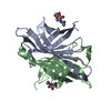 7p4zC  6taaS S: Starting model for refinement C: citing same article ( |
|---|---|
| Similar structure data |


-Links
 PDBj
PDBj

- Assembly
| Deposited unit | 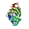
| ||||||||||||
|---|---|---|---|---|---|---|---|---|---|---|---|---|---|
| 1 |
| ||||||||||||
| Unit cell |
|
-Components
| #1: Protein | Mass: 52351.816 Da / Num. of mol.: 1 / Source method: isolated from a natural source / Source: (natural) |
|---|---|
| #2: Polysaccharide | 2-acetamido-2-deoxy-beta-D-glucopyranose-(1-4)-2-acetamido-2-deoxy-beta-D-glucopyranose Source method: isolated from a genetically manipulated source |
| #3: Chemical | ChemComp-CA /   Mass: 40.078 Da / Num. of mol.: 1 / Source method: obtained synthetically / Formula: Ca Mass: 40.078 Da / Num. of mol.: 1 / Source method: obtained synthetically / Formula: Ca |
| #4: Chemical | ChemComp-MES /   Mass: 195.237 Da / Num. of mol.: 1 / Source method: obtained synthetically / Formula: C6H13NO4S / Comment: pH buffer*YM Mass: 195.237 Da / Num. of mol.: 1 / Source method: obtained synthetically / Formula: C6H13NO4S / Comment: pH buffer*YM |
| #5: Water | ChemComp-HOH /  Mass: 18.015 Da / Num. of mol.: 58 / Source method: isolated from a natural source / Formula: H2O Mass: 18.015 Da / Num. of mol.: 58 / Source method: isolated from a natural source / Formula: H2O |
| Has ligand of interest | N |
| Has protein modification | Y |
-Experimental details
-Experiment
| Experiment | Method: X-RAY DIFFRACTION / Number of used crystals: 1 |
|---|
- Sample preparation
| Crystal | Density Matthews: 3.36 Å3/Da / Density % sol: 63.37 % |
|---|---|
| Crystal grow | Temperature: 293 K / Method: vapor diffusion, sitting drop Details: 10% PEG 20k, 20% PEG 500mme, 0.1 M MES/Imidazole pH 6.5, 20 mM of each Polyamine (Morpheus Fusion screen, well H11) |
-Data collection
| Diffraction | Mean temperature: 100 K / Serial crystal experiment: N |
|---|---|
| Diffraction source | Source: SYNCHROTRON / Site: Diamond  / Beamline: I04 / Wavelength: 0.9795 Å / Beamline: I04 / Wavelength: 0.9795 Å |
| Detector | Type: DECTRIS EIGER2 XE 16M / Detector: PIXEL / Date: Jul 2, 2021 |
| Radiation | Protocol: SINGLE WAVELENGTH / Monochromatic (M) / Laue (L): M / Scattering type: x-ray |
| Radiation wavelength | Wavelength: 0.9795 Å / Relative weight: 1 |
| Reflection | Resolution: 2.28→51.92 Å / Num. obs: 32915 / % possible obs: 99.7 % / Redundancy: 9.2 % / Biso Wilson estimate: 36.9 Å2 / CC1/2: 0.99 / Rmerge(I) obs: 9.9 / Rpim(I) all: 0.17 / Rrim(I) all: 4.9 / Net I/σ(I): 9.2 |
| Reflection shell | Resolution: 2.28→2.32 Å / Rmerge(I) obs: 2.27 / Mean I/σ(I) obs: 0.4 / Num. unique obs: 1549 / CC1/2: 0.27 / Rpim(I) all: 1.1 / Rrim(I) all: 2.53 |
- Processing
| Software |
| ||||||||||||||||||||||||||||||||||||||||||||||||||||||||||||||||||||||||||||||||||||
|---|---|---|---|---|---|---|---|---|---|---|---|---|---|---|---|---|---|---|---|---|---|---|---|---|---|---|---|---|---|---|---|---|---|---|---|---|---|---|---|---|---|---|---|---|---|---|---|---|---|---|---|---|---|---|---|---|---|---|---|---|---|---|---|---|---|---|---|---|---|---|---|---|---|---|---|---|---|---|---|---|---|---|---|---|---|
| Refinement | Method to determine structure: MOLECULAR REPLACEMENT Starting model: 6TAA Resolution: 2.28→51.91 Å / SU ML: 0.4488 / Cross valid method: FREE R-VALUE / σ(F): 1.33 / Phase error: 33.3493 Stereochemistry target values: GeoStd + Monomer Library + CDL v1.2
| ||||||||||||||||||||||||||||||||||||||||||||||||||||||||||||||||||||||||||||||||||||
| Solvent computation | Shrinkage radii: 0.9 Å / VDW probe radii: 1.11 Å / Solvent model: FLAT BULK SOLVENT MODEL | ||||||||||||||||||||||||||||||||||||||||||||||||||||||||||||||||||||||||||||||||||||
| Displacement parameters | Biso mean: 53.44 Å2 | ||||||||||||||||||||||||||||||||||||||||||||||||||||||||||||||||||||||||||||||||||||
| Refinement step | Cycle: LAST / Resolution: 2.28→51.91 Å
| ||||||||||||||||||||||||||||||||||||||||||||||||||||||||||||||||||||||||||||||||||||
| Refine LS restraints |
| ||||||||||||||||||||||||||||||||||||||||||||||||||||||||||||||||||||||||||||||||||||
| LS refinement shell |
|
