 Movie
Movie Controller

[English] 日本語
 Yorodumi
- PDB-7p24: Sulfated host glycan recognition by carbohydrate sulfatases of th... -
+ Open data

- Basic information
| Entry | Database: PDB / ID: 7p24 | ||||||
|---|---|---|---|---|---|---|---|
| Title | Sulfated host glycan recognition by carbohydrate sulfatases of the human gut microbiota (BT3177_S1_11) | ||||||
 Components Components | Mucin-desulfating sulfatase | ||||||
 Keywords Keywords | HYDROLASE / Host glycans / sulfation / carbohydrate sulfatases / microbiota | ||||||
| Function / homology |  Function and homology information Function and homology information | ||||||
| Biological species |  Bacteroides thetaiotaomicron (bacteria) Bacteroides thetaiotaomicron (bacteria) | ||||||
| Method |  X-RAY DIFFRACTION / SYNCHROTRON / MOLECULAR REPLACEMENT / Resolution: 0.97 Å X-RAY DIFFRACTION / SYNCHROTRON / MOLECULAR REPLACEMENT / Resolution: 0.97 Å | ||||||
 Authors Authors | Cartmell, A. | ||||||
| Funding support |  United Kingdom, 1items United Kingdom, 1items
| ||||||
 Citation Citation | Journal: Nat.Chem.Biol. / Year: 2022 Title: Sulfated glycan recognition by carbohydrate sulfatases of the human gut microbiota. Authors: Luis, A.S. / Basle, A. / Byrne, D.P. / Wright, G.S.A. / London, J.A. / Jin, C. / Karlsson, N.G. / Hansson, G.C. / Eyers, P.A. / Czjzek, M. / Barbeyron, T. / Yates, E.A. / Martens, E.C. / Cartmell, A. | ||||||
| History |
|
- Structure visualization
| Structure viewer | Molecule: MolmilJmol/JSmol |
|---|
- Downloads & links
-Download
| PDBx/mmCIF format | 7p24.cif.gz | 241.2 KB | Display | PDBx/mmCIF format |
|---|---|---|---|---|
| PDB format | pdb7p24.ent.gz | Display | PDB format | |
| PDBx/mmJSON format | 7p24.json.gz | Tree view | PDBx/mmJSON format | |
| Others |  Other downloads Other downloads |
-Validation report
| Arichive directory | https://data.pdbj.org/pub/pdb/validation_reports/p2/7p24ftp://data.pdbj.org/pub/pdb/validation_reports/p2/7p24 | HTTPS FTP |
|---|
-Related structure data
| Related structure data |  7oz8C 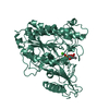 7oz9C 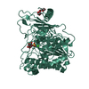 7ozaC 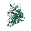 7ozcC 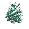 7ozeC 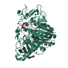 7p26C 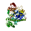 5g2vS S: Starting model for refinement C: citing same article ( |
|---|---|
| Similar structure data |
-Links
 PDBj
PDBj


- Assembly
| Deposited unit | 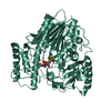
| ||||||||
|---|---|---|---|---|---|---|---|---|---|
| 1 |
| ||||||||
| Unit cell |
|
-Components
| #1: Protein | Mass: 61037.914 Da / Num. of mol.: 1 Source method: isolated from a genetically manipulated source Source: (gene. exp.) Bacteroides thetaiotaomicron (strain ATCC 29148 / DSM 2079 / NCTC 10582 / E50 / VPI-5482) (bacteria)Strain: ATCC 29148 / DSM 2079 / NCTC 10582 / E50 / VPI-5482 / Gene: BT_3177 Production host: References: UniProt: Q8A2X8 |
|---|---|
| #2: Sugar | ChemComp-NGS / 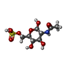  Type: D-saccharide, beta linking / Mass: 301.271 Da / Num. of mol.: 1 / Source method: obtained synthetically / Formula: C8H15NO9S / Feature type: SUBJECT OF INVESTIGATION Type: D-saccharide, beta linking / Mass: 301.271 Da / Num. of mol.: 1 / Source method: obtained synthetically / Formula: C8H15NO9S / Feature type: SUBJECT OF INVESTIGATION |
| #3: Sugar | ChemComp-NGY / 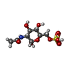  Type: D-saccharide, alpha linking / Mass: 301.271 Da / Num. of mol.: 1 / Source method: obtained synthetically / Formula: C8H15NO9S / Feature type: SUBJECT OF INVESTIGATION Type: D-saccharide, alpha linking / Mass: 301.271 Da / Num. of mol.: 1 / Source method: obtained synthetically / Formula: C8H15NO9S / Feature type: SUBJECT OF INVESTIGATION |
| #4: Chemical | ChemComp-CA /   Mass: 40.078 Da / Num. of mol.: 1 / Source method: obtained synthetically / Formula: Ca Mass: 40.078 Da / Num. of mol.: 1 / Source method: obtained synthetically / Formula: Ca |
| #5: Water | ChemComp-HOH /  Mass: 18.015 Da / Num. of mol.: 593 / Source method: isolated from a natural source / Formula: H2O Mass: 18.015 Da / Num. of mol.: 593 / Source method: isolated from a natural source / Formula: H2O |
| Has ligand of interest | Y |
-Experimental details
-Experiment
| Experiment | Method: X-RAY DIFFRACTION / Number of used crystals: 1 |
|---|
- Sample preparation
| Crystal | Density Matthews: 2.3 Å3/Da / Density % sol: 46.48 % |
|---|---|
| Crystal grow | Temperature: 293 K / Method: vapor diffusion, sitting drop Details: 20 mg/ml BT3177S1_11 with 10 mM 6S-GlcNAc crystallised in 50 % precipitant mix 1 (40% v/v PEG 500, MME; 20 % w/v PEG 20000), 0.1 M carboxcylic acids (0.2M Sodium formate; 0.2M Ammonium ...Details: 20 mg/ml BT3177S1_11 with 10 mM 6S-GlcNAc crystallised in 50 % precipitant mix 1 (40% v/v PEG 500, MME; 20 % w/v PEG 20000), 0.1 M carboxcylic acids (0.2M Sodium formate; 0.2M Ammonium acetate; 0.2M Sodium citrate tribasic dihydrate; 0.2M Sodium potassium tartrate tetrahydrate; 0.2M Sodium oxamate) and 0.1 M buffer system 3 pH 8.5 (Tris (base); BICINE). |
-Data collection
| Diffraction | Mean temperature: 100 K / Serial crystal experiment: N |
|---|---|
| Diffraction source | Source: SYNCHROTRON / Site: Diamond / Beamline: I04-1 / Wavelength: 0.9159 Å |
| Detector | Type: DECTRIS PILATUS 6M-F / Detector: PIXEL / Date: Jul 18, 2018 |
| Radiation | Protocol: SINGLE WAVELENGTH / Monochromatic (M) / Laue (L): M / Scattering type: x-ray |
| Radiation wavelength | Wavelength: 0.9159 Å / Relative weight: 1 |
| Reflection | Resolution: 0.97→35.856 Å / Num. obs: 298423 / % possible obs: 91.9 % / Redundancy: 1.9 % / CC1/2: 0.996 / Net I/σ(I): 8.8 |
| Reflection shell | Resolution: 0.97→0.99 Å / Mean I/σ(I) obs: 1.5 / Num. unique obs: 12666 / CC1/2: 0.596 |
- Processing
| Software |
| |||||||||||||||||||||||||||||||||||||||||||||||||||||||||||||||||||||||||||||||||||||||||||||||||||||||||||||||||||||||||||||||||||||||||||||||||||||||||||||||||||||
|---|---|---|---|---|---|---|---|---|---|---|---|---|---|---|---|---|---|---|---|---|---|---|---|---|---|---|---|---|---|---|---|---|---|---|---|---|---|---|---|---|---|---|---|---|---|---|---|---|---|---|---|---|---|---|---|---|---|---|---|---|---|---|---|---|---|---|---|---|---|---|---|---|---|---|---|---|---|---|---|---|---|---|---|---|---|---|---|---|---|---|---|---|---|---|---|---|---|---|---|---|---|---|---|---|---|---|---|---|---|---|---|---|---|---|---|---|---|---|---|---|---|---|---|---|---|---|---|---|---|---|---|---|---|---|---|---|---|---|---|---|---|---|---|---|---|---|---|---|---|---|---|---|---|---|---|---|---|---|---|---|---|---|---|---|---|---|
| Refinement | Method to determine structure: MOLECULAR REPLACEMENT Starting model: 5G2V Resolution: 0.97→35.856 Å / Cor.coef. Fo:Fc: 0.983 / Cor.coef. Fo:Fc free: 0.981 / WRfactor Rfree: 0.141 / WRfactor Rwork: 0.126 / SU B: 0.705 / SU ML: 0.016 / Average fsc free: 0.9606 / Average fsc work: 0.9634 / Cross valid method: FREE R-VALUE / ESU R: 0.018 / ESU R Free: 0.019 Details: Hydrogens have been added in their riding positions
| |||||||||||||||||||||||||||||||||||||||||||||||||||||||||||||||||||||||||||||||||||||||||||||||||||||||||||||||||||||||||||||||||||||||||||||||||||||||||||||||||||||
| Solvent computation | Ion probe radii: 0.8 Å / Shrinkage radii: 0.8 Å / VDW probe radii: 1.2 Å / Solvent model: MASK BULK SOLVENT | |||||||||||||||||||||||||||||||||||||||||||||||||||||||||||||||||||||||||||||||||||||||||||||||||||||||||||||||||||||||||||||||||||||||||||||||||||||||||||||||||||||
| Displacement parameters | Biso mean: 11.374 Å2
| |||||||||||||||||||||||||||||||||||||||||||||||||||||||||||||||||||||||||||||||||||||||||||||||||||||||||||||||||||||||||||||||||||||||||||||||||||||||||||||||||||||
| Refinement step | Cycle: LAST / Resolution: 0.97→35.856 Å
| |||||||||||||||||||||||||||||||||||||||||||||||||||||||||||||||||||||||||||||||||||||||||||||||||||||||||||||||||||||||||||||||||||||||||||||||||||||||||||||||||||||
| Refine LS restraints |
| |||||||||||||||||||||||||||||||||||||||||||||||||||||||||||||||||||||||||||||||||||||||||||||||||||||||||||||||||||||||||||||||||||||||||||||||||||||||||||||||||||||
| LS refinement shell |
|
