- PDB-7p0s: ORF virus encoded Bcl-2 homolog ORFV125 in complex with Puma BH3 ... -
+
Open data
ID or keywords:
Loading...
-
Basic information
Entry
Database: PDB / ID: 7p0s
Title
ORF virus encoded Bcl-2 homolog ORFV125 in complex with Puma BH3 peptide
Components
Apoptosis inhibitor
Bcl-2-binding component 3, isoforms 1/2
Keywords
APOPTOSIS / ORF virus / Bcl-2
Function / homology
Function and homology information
positive regulation of establishment of protein localization to mitochondrion / positive regulation of endoplasmic reticulum stress-induced intrinsic apoptotic signaling pathway / positive regulation of fibroblast apoptotic process / T cell apoptotic process / BH3-only proteins associate with and inactivate anti-apoptotic BCL-2 members / positive regulation of thymocyte apoptotic process / fibroblast apoptotic process / Activation of PUMA and translocation to mitochondria / execution phase of apoptosis / FOXO-mediated transcription of cell death genes ...positive regulation of establishment of protein localization to mitochondrion / positive regulation of endoplasmic reticulum stress-induced intrinsic apoptotic signaling pathway / positive regulation of fibroblast apoptotic process / T cell apoptotic process / BH3-only proteins associate with and inactivate anti-apoptotic BCL-2 members / positive regulation of thymocyte apoptotic process / fibroblast apoptotic process / Activation of PUMA and translocation to mitochondria / execution phase of apoptosis / FOXO-mediated transcription of cell death genes / positive regulation of IRE1-mediated unfolded protein response / positive regulation of release of cytochrome c from mitochondria / TP53 Regulates Transcription of Genes Involved in Cytochrome C Release / intrinsic apoptotic signaling pathway in response to endoplasmic reticulum stress / intrinsic apoptotic signaling pathway in response to DNA damage by p53 class mediator / positive regulation of intrinsic apoptotic signaling pathway / intrinsic apoptotic signaling pathway / response to endoplasmic reticulum stress / release of cytochrome c from mitochondria / determination of adult lifespan / cellular response to ionizing radiation / apoptotic signaling pathway / positive regulation of protein-containing complex assembly / positive regulation of neuron apoptotic process / cellular response to hypoxia / mitochondrial outer membrane / DNA damage response / mitochondrion / metal ion binding / membrane / cytosol Similarity search - Function
In the structure databanks used in Yorodumi, some data are registered as the other names, "COVID-19 virus" and "2019-nCoV". Here are the details of the virus and the list of structure data.
Jan 31, 2019. EMDB accession codes are about to change! (news from PDBe EMDB page)
EMDB accession codes are about to change! (news from PDBe EMDB page)
The allocation of 4 digits for EMDB accession codes will soon come to an end. Whilst these codes will remain in use, new EMDB accession codes will include an additional digit and will expand incrementally as the available range of codes is exhausted. The current 4-digit format prefixed with “EMD-” (i.e. EMD-XXXX) will advance to a 5-digit format (i.e. EMD-XXXXX), and so on. It is currently estimated that the 4-digit codes will be depleted around Spring 2019, at which point the 5-digit format will come into force.
The EM Navigator/Yorodumi systems omit the EMD- prefix.
Related info.:Q: What is EMD? / ID/Accession-code notation in Yorodumi/EM Navigator
Yorodumi is a browser for structure data from EMDB, PDB, SASBDB, etc.
This page is also the successor to EM Navigator detail page, and also detail information page/front-end page for Omokage search.
The word "yorodu" (or yorozu) is an old Japanese word meaning "ten thousand". "mi" (miru) is to see.
Related info.:EMDB / PDB / SASBDB / Comparison of 3 databanks / Yorodumi Search / Aug 31, 2016. New EM Navigator & Yorodumi / Yorodumi Papers / Jmol/JSmol / Function and homology information / Changes in new EM Navigator and Yorodumi
 Movie
Movie Controller

 Yorodumi
 Open data
 Basic information
 Components
 Keywords
 Function and homology information
 Orf virus
 Homo sapiens (human)
 X-RAY DIFFRACTION /
  Authors
 Australia, 1items
  Citation
 Structure visualization
 Downloads & links
 Other downloads
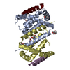

 PDBj
PDBj


 Assembly
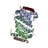

 Mass: 18.015 Da / Num. of mol.: 25 / Source method: isolated from a natural source / Formula: H2O
 Sample preparation
 Processing