| Entry | Database: PDB / ID: 7p0o
|
|---|
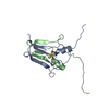
| Title | mitoNEET bound to M1 molecule |
|---|
 Components Components | CDGSH iron-sulfur domain-containing protein 1 |
|---|
 Keywords Keywords | METAL BINDING PROTEIN / [2Fe-2S] proteins / NEET proteins / destabilizer / M1 |
|---|
| Function / homology |  Function and homology information Function and homology information
cysteine transaminase / L-cysteine:2-oxoglutarate transaminase activity / regulation of cellular respiration / regulation of autophagy / protein maturation / 2 iron, 2 sulfur cluster binding / pyridoxal phosphate binding / intracellular iron ion homeostasis / mitochondrial outer membrane / protein homodimerization activity ...cysteine transaminase / L-cysteine:2-oxoglutarate transaminase activity / regulation of cellular respiration / regulation of autophagy / protein maturation / 2 iron, 2 sulfur cluster binding / pyridoxal phosphate binding / intracellular iron ion homeostasis / mitochondrial outer membrane / protein homodimerization activity / mitochondrion / metal ion binding / identical protein bindingSimilarity search - Function Iron sulphur domain-containing, mitoNEET, N-terminal / Iron-containing outer mitochondrial membrane protein N-terminus / CDGSH iron-sulfur domain-containing protein 1/2 / Iron-binding zinc finger CDGSH type / Iron-binding zinc finger, CDGSH type / MitoNEET, CDGSH iron-sulfur domain / CDGSH-type zinc finger. Function unknown.Similarity search - Domain/homology |
|---|
| Biological species |  Homo sapiens (human) Homo sapiens (human) |
|---|
| Method |  X-RAY DIFFRACTION / SYNCHROTRON / MOLECULAR REPLACEMENT / Resolution: 1.65 Å X-RAY DIFFRACTION / SYNCHROTRON / MOLECULAR REPLACEMENT / Resolution: 1.65 Å |
|---|
 Authors Authors | Livnah, O. / Eisenberg-Domovich, Y. / Marjault, H.B. / Nechushtai, R. |
|---|
| Funding support |  Israel, 1items Israel, 1items | Organization | Grant number | Country |
|---|
| Marie Sklodowska-Curie Actions, FragNET ITN | 765048 | Israel |
|
|---|
 Citation Citation | Journal: Commun Biol / Year: 2022
Title: An anti-diabetic drug targets NEET (CISD) proteins through destabilization of their [2Fe-2S] clusters.
Authors: Marjault, H.B. / Karmi, O. / Zuo, K. / Michaeli, D. / Eisenberg-Domovich, Y. / Rossetti, G. / de Chassey, B. / Vonderscher, J. / Cabantchik, I. / Carloni, P. / Mittler, R. / Livnah, O. / ...Authors: Marjault, H.B. / Karmi, O. / Zuo, K. / Michaeli, D. / Eisenberg-Domovich, Y. / Rossetti, G. / de Chassey, B. / Vonderscher, J. / Cabantchik, I. / Carloni, P. / Mittler, R. / Livnah, O. / Meldrum, E. / Nechushtai, R. |
|---|
| History | | Deposition | Jun 30, 2021 | Deposition site: PDBE / Processing site: PDBE |
|---|
| Revision 1.0 | May 25, 2022 | Provider: repository / Type: Initial release |
|---|
| Revision 1.1 | Jan 31, 2024 | Group: Data collection / Refinement description
Category: chem_comp_atom / chem_comp_bond / pdbx_initial_refinement_model |
|---|
|
|---|
 Movie
Movie Controller


 Open data
 Basic information
 Structure visualization
 Downloads & links
 Other downloads
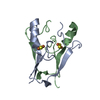
 PDBj
PDBj


 Assembly
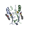


 Mass: 175.820 Da / Num. of mol.: 2 / Source method: obtained synthetically / Formula: Fe2S2
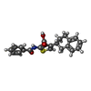
 Mass: 377.456 Da / Num. of mol.: 1 / Source method: obtained synthetically / Formula: C22H19NO3S / Feature type: SUBJECT OF INVESTIGATION
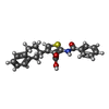
 Mass: 377.456 Da / Num. of mol.: 1 / Source method: obtained synthetically / Formula: C22H19NO3S
 Mass: 18.015 Da / Num. of mol.: 50 / Source method: isolated from a natural source / Formula: H2O
 Sample preparation
 / Beamline: 14.1 / Wavelength: 0.9184 Å
 Processing