[English] 日本語
Yorodumi
- PDB-7oxx: CrabP2 mutant R30AK31A -

+
Open data


ID or keywords:

Loading...

-
Basic information

Entry
Database: PDB / ID: 7oxx
TitleCrabP2 mutant R30AK31A
ComponentsCellular retinoic acid-binding protein 2
KeywordsTRANSPORT PROTEIN / retinoic acid / CRABP2 / cyclin / CDK4/6 / nuclear hormone receptor / signalling kinase
Function / homology
Function and homology information


positive regulation of collateral sprouting / retinoid binding / retinal binding / retinoic acid binding / embryonic forelimb morphogenesis / retinoic acid metabolic process / retinol binding / Signaling by Retinoic Acid / epidermis development / fatty acid transport ...positive regulation of collateral sprouting / retinoid binding / retinal binding / retinoic acid binding / embryonic forelimb morphogenesis / retinoic acid metabolic process / retinol binding / Signaling by Retinoic Acid / epidermis development / fatty acid transport / cyclin binding / fatty acid binding / regulation of DNA-templated transcription / endoplasmic reticulum / signal transduction / extracellular exosome / nucleoplasm / nucleus / cytosol / cytoplasm
Similarity search - Function
Cytosolic fatty-acid binding proteins signature. / Intracellular lipid binding protein / Cytosolic fatty-acid binding / Lipocalin / cytosolic fatty-acid binding protein family / Lipocalin/cytosolic fatty-acid binding domain / Calycin
Similarity search - Domain/homology
Cellular retinoic acid-binding protein 2
Similarity search - Component
Biological speciesHomo sapiens (human)
MethodX-RAY DIFFRACTION / SYNCHROTRON / MOLECULAR REPLACEMENT / Resolution: 1.33 Å
AuthorsTomlinson, C.W.E. / Basle, A. / Pohl, E.
Funding support United Kingdom, 1items
OrganizationGrant numberCountry
Medical Research Council (MRC, United Kingdom)MR/N009738/1 United Kingdom
CitationJournal: To Be Published
Title: Structural requirements for the specific binding of CRABP2 to cyclin D3
Authors: Pastok, M.W. / Tomlinson, C.W.E. / Tatum, N.J. / Basle, A. / Noble, M.E.M. / Pohl, E. / Endicott, J.A.
History
DepositionJun 23, 2021Deposition site: PDBE / Processing site: PDBE
Revision 1.0Jul 13, 2022Provider: repository / Type: Initial release
Revision 1.1Jan 31, 2024Group: Data collection / Refinement description
Category: chem_comp_atom / chem_comp_bond ...chem_comp_atom / chem_comp_bond / pdbx_initial_refinement_model / struct_ncs_dom_lim
Item: _struct_ncs_dom_lim.beg_auth_comp_id / _struct_ncs_dom_lim.beg_label_asym_id ..._struct_ncs_dom_lim.beg_auth_comp_id / _struct_ncs_dom_lim.beg_label_asym_id / _struct_ncs_dom_lim.beg_label_comp_id / _struct_ncs_dom_lim.beg_label_seq_id / _struct_ncs_dom_lim.end_auth_comp_id / _struct_ncs_dom_lim.end_label_asym_id / _struct_ncs_dom_lim.end_label_comp_id / _struct_ncs_dom_lim.end_label_seq_id

-
Structure visualization

Structure viewerMolecule:
MolmilJmol/JSmol

Downloads & links

-
Assembly

Deposited unit
A: Cellular retinoic acid-binding protein 2
B: Cellular retinoic acid-binding protein 2
C: Cellular retinoic acid-binding protein 2
D: Cellular retinoic acid-binding protein 2
hetero molecules


Theoretical massNumber of molelcules
Total (without water)62,3678
Polymers62,2754
Non-polymers924
Water7,188399
1


  • Idetical with deposited unit
  • defined by author&software
TypeNameSymmetry operationNumber
identity operation1_555x,y,z1
Buried area17290 Å2
ΔGint-161 kcal/mol
Surface area24800 Å2
MethodPISA
Unit cell
Length a, b, c (Å)51.170, 106.020, 55.540
Angle α, β, γ (deg.)90.000, 101.700, 90.000
Int Tables number4
Space group name H-MP1211
Noncrystallographic symmetry (NCS)NCS domain:
IDEns-IDDetails
11A
21B
32A
42C
53A
63D
74B
84C
95B
105D
116C
126D

NCS domain segments:
Dom-IDComponent-IDEns-IDBeg auth comp-IDBeg label comp-IDEnd auth comp-IDEnd label comp-IDAuth asym-IDLabel asym-IDAuth seq-IDLabel seq-ID
111ASNASNARGARGAA3 - 1373 - 137
221ASNASNARGARGBB3 - 1373 - 137
332ASNASNARGARGAA3 - 1373 - 137
442ASNASNARGARGCC3 - 1373 - 137
553ASNASNARGARGAA3 - 1373 - 137
663ASNASNARGARGDD3 - 1373 - 137
774PROPROARGARGBB2 - 1372 - 137
884PROPROARGARGCC2 - 1372 - 137
995PROPROARGARGBB2 - 1372 - 137
10105PROPROARGARGDD2 - 1372 - 137
11116METMETGLUGLUCC1 - 1381 - 138
12126METMETGLUGLUDD1 - 1381 - 138

NCS ensembles :
IDDetails
1Local NCS retraints between domains: 1 2
2Local NCS retraints between domains: 3 4
3Local NCS retraints between domains: 5 6
4Local NCS retraints between domains: 7 8
5Local NCS retraints between domains: 9 10
6Local NCS retraints between domains: 11 12

-
Components

#1: Protein
Cellular retinoic acid-binding protein 2 / Cellular retinoic acid-binding protein II / CRABP-II


Mass: 15568.781 Da / Num. of mol.: 4
Source method: isolated from a genetically manipulated source
Source: (gene. exp.) Homo sapiens (human) / Gene: CRABP2 / Production host: Escherichia coli BL21(DE3) (bacteria) / References: UniProt: P29373
#2: Chemical
ChemComp-NA / SODIUM ION


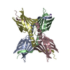
Mass: 22.990 Da / Num. of mol.: 4 / Source method: obtained synthetically / Formula: Na
#3: Water ChemComp-HOH / water / Water


Mass: 18.015 Da / Num. of mol.: 399 / Source method: isolated from a natural source / Formula: H2O
Has ligand of interestN

-
Experimental details

-
Experiment

ExperimentMethod: X-RAY DIFFRACTION / Number of used crystals: 1

-
Sample preparation

CrystalDensity Matthews: 2.37 Å3/Da / Density % sol: 48.08 %
Crystal growTemperature: 293.15 K / Method: vapor diffusion, sitting drop
Details: 150 mM MgCl2, 100 mM Bis-Tris pH 6.5 and 25% PEG 3350.

-
Data collection

DiffractionMean temperature: 100 K / Serial crystal experiment: N
Diffraction sourceSource: SYNCHROTRON / Site: Diamond / Beamline: I24 / Wavelength: 0.9686 Å
DetectorType: DECTRIS PILATUS 6M / Detector: PIXEL / Date: Dec 8, 2019
RadiationProtocol: SINGLE WAVELENGTH / Monochromatic (M) / Laue (L): M / Scattering type: x-ray
Radiation wavelengthWavelength: 0.9686 Å / Relative weight: 1
ReflectionResolution: 1.33→54.39 Å / Num. obs: 128510 / % possible obs: 97.1 % / Redundancy: 3.3 % / CC1/2: 0.996 / Net I/σ(I): 9.7
Reflection shellResolution: 1.33→1.35 Å / Redundancy: 3.1 % / Mean I/σ(I) obs: 1.4 / Num. unique obs: 6247 / CC1/2: 0.411 / % possible all: 94.7

-
Processing

Software
NameVersionClassification
REFMAC5.8.0267refinement
xia2data reduction
XDSdata reduction
Aimlessdata scaling
PHASERphasing
Cootmodel building
MolProbitymodel building
RefinementMethod to determine structure: MOLECULAR REPLACEMENT
Starting model: 2FRS
Resolution: 1.33→54.386 Å / Cor.coef. Fo:Fc: 0.975 / Cor.coef. Fo:Fc free: 0.963 / SU B: 3.376 / SU ML: 0.057 / Cross valid method: FREE R-VALUE / ESU R: 0.054 / ESU R Free: 0.054
Details: Hydrogens have been added in their riding positions
RfactorNum. reflection% reflection
Rfree0.1996 6503 5.062 %
Rwork0.1584 121967 -
all0.161 --
obs-128470 96.888 %
Solvent computationIon probe radii: 0.8 Å / Shrinkage radii: 0.8 Å / VDW probe radii: 1.2 Å / Solvent model: MASK BULK SOLVENT
Displacement parametersBiso mean: 25.357 Å2
Baniso -1Baniso -2Baniso -3
1-3.599 Å20 Å21.37 Å2
2---3.617 Å20 Å2
3----0.506 Å2
Refinement stepCycle: LAST / Resolution: 1.33→54.386 Å
ProteinNucleic acidLigandSolventTotal
Num. atoms4333 0 4 399 4736
Refine LS restraints
Refine-IDTypeDev idealDev ideal targetNumber
X-RAY DIFFRACTIONr_bond_refined_d0.0150.0134532
X-RAY DIFFRACTIONr_bond_other_d0.0120.0174438
X-RAY DIFFRACTIONr_angle_refined_deg1.8591.6516158
X-RAY DIFFRACTIONr_angle_other_deg1.5871.58110284
X-RAY DIFFRACTIONr_dihedral_angle_1_deg7.9255589
X-RAY DIFFRACTIONr_dihedral_angle_2_deg33.94223.604222
X-RAY DIFFRACTIONr_dihedral_angle_3_deg12.80815877
X-RAY DIFFRACTIONr_dihedral_angle_4_deg11.0261525
X-RAY DIFFRACTIONr_chiral_restr0.1180.2623
X-RAY DIFFRACTIONr_gen_planes_refined0.010.025087
X-RAY DIFFRACTIONr_gen_planes_other0.0060.02975
X-RAY DIFFRACTIONr_nbd_refined0.1950.2634
X-RAY DIFFRACTIONr_symmetry_nbd_other0.1920.24053
X-RAY DIFFRACTIONr_nbtor_refined0.1560.22088
X-RAY DIFFRACTIONr_symmetry_nbtor_other0.0760.22300
X-RAY DIFFRACTIONr_xyhbond_nbd_refined0.1340.2269
X-RAY DIFFRACTIONr_symmetry_xyhbond_nbd_other0.0360.22
X-RAY DIFFRACTIONr_metal_ion_refined0.3390.212
X-RAY DIFFRACTIONr_symmetry_nbd_refined0.1130.26
X-RAY DIFFRACTIONr_nbd_other0.1410.226
X-RAY DIFFRACTIONr_symmetry_xyhbond_nbd_refined0.1680.225
X-RAY DIFFRACTIONr_mcbond_it2.6992.2352239
X-RAY DIFFRACTIONr_mcbond_other2.5572.2332238
X-RAY DIFFRACTIONr_mcangle_it3.2183.3742804
X-RAY DIFFRACTIONr_mcangle_other3.2363.3762805
X-RAY DIFFRACTIONr_scbond_it4.1352.8142293
X-RAY DIFFRACTIONr_scbond_other4.1072.812290
X-RAY DIFFRACTIONr_scangle_it4.9974.0033333
X-RAY DIFFRACTIONr_scangle_other4.9854.0013332
X-RAY DIFFRACTIONr_lrange_it4.59826.5144807
X-RAY DIFFRACTIONr_lrange_other4.52426.1844735
X-RAY DIFFRACTIONr_rigid_bond_restr8.60238970
X-RAY DIFFRACTIONr_ncsr_local_group_10.0790.053929
X-RAY DIFFRACTIONr_ncsr_local_group_20.1040.053850
X-RAY DIFFRACTIONr_ncsr_local_group_30.1110.053830
X-RAY DIFFRACTIONr_ncsr_local_group_40.1080.053842
X-RAY DIFFRACTIONr_ncsr_local_group_50.1130.053802
X-RAY DIFFRACTIONr_ncsr_local_group_60.0990.053865
Refine LS restraints NCS
Ens-IDDom-IDAuth asym-IDRefine-IDTypeRms dev position (Å)Weight position
11AX-RAY DIFFRACTIONLocal ncs0.078910.05008
12BX-RAY DIFFRACTIONLocal ncs0.078910.05008
23AX-RAY DIFFRACTIONLocal ncs0.104460.05008
24CX-RAY DIFFRACTIONLocal ncs0.104460.05008
35AX-RAY DIFFRACTIONLocal ncs0.111270.05008
36DX-RAY DIFFRACTIONLocal ncs0.111270.05008
47BX-RAY DIFFRACTIONLocal ncs0.108460.05008
48CX-RAY DIFFRACTIONLocal ncs0.108460.05008
59BX-RAY DIFFRACTIONLocal ncs0.113060.05008
510DX-RAY DIFFRACTIONLocal ncs0.113060.05008
611CX-RAY DIFFRACTIONLocal ncs0.098640.05008
612DX-RAY DIFFRACTIONLocal ncs0.098640.05008
LS refinement shell

Refine-ID: X-RAY DIFFRACTION / Total num. of bins used: 20

Resolution (Å)Rfactor RfreeNum. reflection RfreeRfactor RworkNum. reflection RworkRfactor allNum. reflection allFsc freeFsc work% reflection obs (%)WRfactor Rwork
1.33-1.3650.3224880.29288180.29497860.7890.83995.0950.278
1.365-1.4020.3084330.26987080.27195300.840.86695.91820.248
1.402-1.4430.2754350.23785230.23892830.8740.89996.4990.214
1.443-1.4870.2364270.20682130.20789780.8980.91596.23520.181
1.487-1.5360.2424350.17580010.17887830.920.94196.04920.152
1.536-1.5890.1954360.1676740.16284410.9470.95396.07870.14
1.589-1.6490.2033840.14375370.14681520.9490.96297.16630.126
1.649-1.7170.1963720.12172630.12478060.9520.97697.80940.109
1.717-1.7930.1913640.13269890.13575510.9590.96897.37780.124
1.793-1.880.1813660.12266140.12571790.9480.9797.2280.119
1.88-1.9820.1843300.12463460.12768720.9550.97297.14780.127
1.982-2.1020.192960.14459810.14664530.950.96197.27260.153
2.102-2.2470.182700.13956990.14161150.9530.96397.61240.153
2.247-2.4270.1862810.14352530.14556750.9530.9697.51540.164
2.427-2.6580.1972580.14448410.14752220.9520.96597.64460.17
2.658-2.970.1862730.15143230.15347200.9520.96197.37290.181
2.97-3.4280.2132460.16838690.17142020.9430.95797.92960.206
3.428-4.1950.1771690.16133120.16235440.9620.96398.22230.199
4.195-5.9140.1981510.16725640.16827520.9650.96998.65550.221
5.914-54.3860.223890.20114400.20215530.9530.95298.45460.268

+
About Yorodumi

-
News

-
Feb 9, 2022. New format data for meta-information of EMDB entries

New format data for meta-information of EMDB entries

  • Version 3 of the EMDB header file is now the official format.
  • The previous official version 1.9 will be removed from the archive.

Related info.:EMDB header

External links:wwPDB to switch to version 3 of the EMDB data model

-
Aug 12, 2020. Covid-19 info

Covid-19 info

URL: https://pdbj.org/emnavi/covid19.php

New page: Covid-19 featured information page in EM Navigator.

Related info.:Covid-19 info / Mar 5, 2020. Novel coronavirus structure data

+
Mar 5, 2020. Novel coronavirus structure data

Novel coronavirus structure data

Related info.:Yorodumi Speices / Aug 12, 2020. Covid-19 info

External links:COVID-19 featured content - PDBj / Molecule of the Month (242):Coronavirus Proteases

+
Jan 31, 2019. EMDB accession codes are about to change! (news from PDBe EMDB page)

EMDB accession codes are about to change! (news from PDBe EMDB page)

  • The allocation of 4 digits for EMDB accession codes will soon come to an end. Whilst these codes will remain in use, new EMDB accession codes will include an additional digit and will expand incrementally as the available range of codes is exhausted. The current 4-digit format prefixed with “EMD-” (i.e. EMD-XXXX) will advance to a 5-digit format (i.e. EMD-XXXXX), and so on. It is currently estimated that the 4-digit codes will be depleted around Spring 2019, at which point the 5-digit format will come into force.
  • The EM Navigator/Yorodumi systems omit the EMD- prefix.

Related info.:Q: What is EMD? / ID/Accession-code notation in Yorodumi/EM Navigator

External links:EMDB Accession Codes are Changing Soon! / Contact to PDBj

+
Jul 12, 2017. Major update of PDB

Major update of PDB

  • wwPDB released updated PDB data conforming to the new PDBx/mmCIF dictionary.
  • This is a major update changing the version number from 4 to 5, and with Remediation, in which all the entries are updated.
  • In this update, many items about electron microscopy experimental information are reorganized (e.g. em_software).
  • Now, EM Navigator and Yorodumi are based on the updated data.

External links:wwPDB Remediation / Enriched Model Files Conforming to OneDep Data Standards Now Available in the PDB FTP Archive

-
Yorodumi

Thousand views of thousand structures

  • Yorodumi is a browser for structure data from EMDB, PDB, SASBDB, etc.
  • This page is also the successor to EM Navigator detail page, and also detail information page/front-end page for Omokage search.
  • The word "yorodu" (or yorozu) is an old Japanese word meaning "ten thousand". "mi" (miru) is to see.

Related info.:EMDB / PDB / SASBDB / Comparison of 3 databanks / Yorodumi Search / Aug 31, 2016. New EM Navigator & Yorodumi / Yorodumi Papers / Jmol/JSmol / Function and homology information / Changes in new EM Navigator and Yorodumi

Read more