 Movie
Movie Controller

[English] 日本語
 Yorodumi
- PDB-7owm: HsNMT1 in complex with both MyrCoA and HCPA substrate peptide GKQNSKLR -
+ Open data

- Basic information
| Entry | Database: PDB / ID: 7owm | ||||||||||||
|---|---|---|---|---|---|---|---|---|---|---|---|---|---|
| Title | HsNMT1 in complex with both MyrCoA and HCPA substrate peptide GKQNSKLR | ||||||||||||
 Components Components |
| ||||||||||||
 Keywords Keywords | TRANSFERASE / E-MYRISTOYLATION / NMT / MYRISTOYLTRANSFERASE TYPE1 / ACYLTRANSFERASE / GNAT / GCN5-RELATED N-ACETYLTRANSFERASES | ||||||||||||
| Function / homology |  Function and homology information Function and homology informationregulation of voltage-gated calcium channel activity / response to Aroclor 1254 / response to ketamine / myristoyltransferase activity / N-terminal peptidyl-glycine N-myristoylation / peptidyl-lysine N6-myristoyltransferase activity / Late Phase of HIV Life Cycle / ketone metabolic process / regulation of opsin-mediated signaling pathway / cellular response to electrical stimulus ...regulation of voltage-gated calcium channel activity / response to Aroclor 1254 / response to ketamine / myristoyltransferase activity / N-terminal peptidyl-glycine N-myristoylation / peptidyl-lysine N6-myristoyltransferase activity / Late Phase of HIV Life Cycle / ketone metabolic process / regulation of opsin-mediated signaling pathway / cellular response to electrical stimulus / Activation, myristolyation of BID and translocation to mitochondria / positive regulation of protein localization to mitochondrion / glycylpeptide N-tetradecanoyltransferase / glycylpeptide N-tetradecanoyltransferase activity / cellular response to L-glutamate / protein localization to membrane / dendritic spine head / regulation of postsynaptic neurotransmitter receptor internalization / neuronal cell body membrane / inner ear development / regulation of signal transduction / positive regulation of protein targeting to membrane / eNOS activation / Transferases; Acyltransferases; Transferring groups other than aminoacyl groups / cellular response to calcium ion / dendrite cytoplasm / dendrite membrane / calcium-mediated signaling / kinase binding / Inactivation, recovery and regulation of the phototransduction cascade / actin binding / retina development in camera-type eye / in utero embryonic development / perikaryon / axon / calcium ion binding / glutamatergic synapse / membrane / identical protein binding / plasma membrane / cytoplasm / cytosol Similarity search - Function | ||||||||||||
| Biological species |  Homo sapiens (human) Homo sapiens (human) | ||||||||||||
| Method |  X-RAY DIFFRACTION / SYNCHROTRON / MOLECULAR REPLACEMENT / Resolution: 1.5 Å X-RAY DIFFRACTION / SYNCHROTRON / MOLECULAR REPLACEMENT / Resolution: 1.5 Å | ||||||||||||
 Authors Authors | Dian, C. / Giglione, C. / Meinnel, T. | ||||||||||||
| Funding support |  France, 3items France, 3items
| ||||||||||||
 Citation Citation | Journal: J.Mol.Biol. / Year: 2022 Title: Structural and Large-scale Analysis Unveil the Intertwined Paths Promoting NMT-catalyzed Lysine and Glycine Myristoylation. Authors: Riviere, F. / Dian, C. / Dutheil, R.F. / Monassa, P. / Giglione, C. / Meinnel, T. | ||||||||||||
| History |
|
- Structure visualization
| Structure viewer | Molecule: MolmilJmol/JSmol |
|---|
- Downloads & links
-Download
| PDBx/mmCIF format | 7owm.cif.gz | 434.8 KB | Display | PDBx/mmCIF format |
|---|---|---|---|---|
| PDB format | pdb7owm.ent.gz | 294.5 KB | Display | PDB format |
| PDBx/mmJSON format | 7owm.json.gz | Tree view | PDBx/mmJSON format | |
| Others |  Other downloads Other downloads |
-Validation report
| Arichive directory | https://data.pdbj.org/pub/pdb/validation_reports/ow/7owmftp://data.pdbj.org/pub/pdb/validation_reports/ow/7owm | HTTPS FTP |
|---|
-Related structure data
| Related structure data |  7ownC  7owoC  7owpC  7owqC 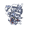 7owrC 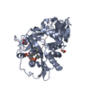 7owuC 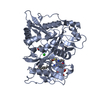 5o9tS S: Starting model for refinement C: citing same article ( |
|---|---|
| Similar structure data |
-Links
 PDBj
PDBj


- Assembly
| Deposited unit | 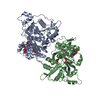
| ||||||||||||
|---|---|---|---|---|---|---|---|---|---|---|---|---|---|
| 1 | 
| ||||||||||||
| 2 | 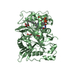
| ||||||||||||
| Unit cell |
|
-Components
| #1: Protein | Mass: 46465.414 Da / Num. of mol.: 2 Source method: isolated from a genetically manipulated source Source: (gene. exp.) Homo sapiens (human) / Gene: NMT1, NMT / Plasmid: pET28 / Production host:  References: UniProt: P30419, glycylpeptide N-tetradecanoyltransferase #2: Protein/peptide | | Mass: 933.088 Da / Num. of mol.: 1 / Source method: obtained synthetically / Details: synthetic peptide / Source: (synth.) Homo sapiens (human) / References: UniProt: P84074#3: Chemical | ChemComp-GOL /   Mass: 92.094 Da / Num. of mol.: 4 / Source method: obtained synthetically / Formula: C3H8O3 Mass: 92.094 Da / Num. of mol.: 4 / Source method: obtained synthetically / Formula: C3H8O3#4: Chemical |   Mass: 977.890 Da / Num. of mol.: 2 / Source method: obtained synthetically / Formula: C35H62N7O17P3S / Feature type: SUBJECT OF INVESTIGATION Mass: 977.890 Da / Num. of mol.: 2 / Source method: obtained synthetically / Formula: C35H62N7O17P3S / Feature type: SUBJECT OF INVESTIGATION#5: Water | ChemComp-HOH / |  Mass: 18.015 Da / Num. of mol.: 1006 / Source method: isolated from a natural source / Formula: H2O Mass: 18.015 Da / Num. of mol.: 1006 / Source method: isolated from a natural source / Formula: H2OHas ligand of interest | Y | Has protein modification | Y | |
|---|
-Experimental details
-Experiment
| Experiment | Method: X-RAY DIFFRACTION / Number of used crystals: 1 |
|---|
- Sample preparation
| Crystal | Density Matthews: 2.31 Å3/Da / Density % sol: 46.69 % |
|---|---|
| Crystal grow | Temperature: 293 K / Method: vapor diffusion, hanging drop / pH: 5.6 Details: 20% PEG8K, 100mM Sodium Citrate pH 5.6, 100mM MgCl2, 100mM NaCl |
-Data collection
| Diffraction | Mean temperature: 100 K / Ambient temp details: Cryostream / Serial crystal experiment: N |
|---|---|
| Diffraction source | Source: SYNCHROTRON / Site: SOLEIL / Beamline: PROXIMA 1 / Wavelength: 0.984 Å |
| Detector | Type: DECTRIS EIGER X 16M / Detector: PIXEL / Date: Jul 18, 2019 / Details: K/B mirrors |
| Radiation | Monochromator: silicium Si(111) / Protocol: SINGLE WAVELENGTH / Monochromatic (M) / Laue (L): M / Scattering type: x-ray |
| Radiation wavelength | Wavelength: 0.984 Å / Relative weight: 1 |
| Reflection | Resolution: 1.5→48.773 Å / Num. obs: 128545 / % possible obs: 98.4 % / Redundancy: 13.9 % / Biso Wilson estimate: 16.7 Å2 / CC1/2: 0.9978 / Rmerge(I) obs: 0.115 / Rpim(I) all: 0.032 / Net I/σ(I): 12.2 |
| Reflection shell | Resolution: 1.5→1.54 Å / Redundancy: 8.9 % / Rmerge(I) obs: 1.532 / Mean I/σ(I) obs: 1.4 / Num. unique obs: 6425 / CC1/2: 0.6483 / Rpim(I) all: 0.541 / % possible all: 97.8 |
- Processing
| Software |
| |||||||||||||||||||||||||||||||||||||||||||||||||||||||||||||||||||||||||||||||||||||||||||||||||||||||||||||||||||||||||||||||||||||||||||||||||||||||||||||||||||||||||||||||||||||||||||||||||||||||||||||||||||||||||
|---|---|---|---|---|---|---|---|---|---|---|---|---|---|---|---|---|---|---|---|---|---|---|---|---|---|---|---|---|---|---|---|---|---|---|---|---|---|---|---|---|---|---|---|---|---|---|---|---|---|---|---|---|---|---|---|---|---|---|---|---|---|---|---|---|---|---|---|---|---|---|---|---|---|---|---|---|---|---|---|---|---|---|---|---|---|---|---|---|---|---|---|---|---|---|---|---|---|---|---|---|---|---|---|---|---|---|---|---|---|---|---|---|---|---|---|---|---|---|---|---|---|---|---|---|---|---|---|---|---|---|---|---|---|---|---|---|---|---|---|---|---|---|---|---|---|---|---|---|---|---|---|---|---|---|---|---|---|---|---|---|---|---|---|---|---|---|---|---|---|---|---|---|---|---|---|---|---|---|---|---|---|---|---|---|---|---|---|---|---|---|---|---|---|---|---|---|---|---|---|---|---|---|---|---|---|---|---|---|---|---|---|---|---|---|---|---|---|---|
| Refinement | Method to determine structure: MOLECULAR REPLACEMENT Starting model: 5O9T Resolution: 1.5→48.77 Å / SU ML: 0.1589 / Cross valid method: FREE R-VALUE / σ(F): 1.35 / Phase error: 19.1827 Stereochemistry target values: GeoStd + Monomer Library + CDL v1.2
| |||||||||||||||||||||||||||||||||||||||||||||||||||||||||||||||||||||||||||||||||||||||||||||||||||||||||||||||||||||||||||||||||||||||||||||||||||||||||||||||||||||||||||||||||||||||||||||||||||||||||||||||||||||||||
| Solvent computation | Shrinkage radii: 0.9 Å / VDW probe radii: 1.11 Å / Solvent model: FLAT BULK SOLVENT MODEL | |||||||||||||||||||||||||||||||||||||||||||||||||||||||||||||||||||||||||||||||||||||||||||||||||||||||||||||||||||||||||||||||||||||||||||||||||||||||||||||||||||||||||||||||||||||||||||||||||||||||||||||||||||||||||
| Displacement parameters | Biso mean: 25.18 Å2 | |||||||||||||||||||||||||||||||||||||||||||||||||||||||||||||||||||||||||||||||||||||||||||||||||||||||||||||||||||||||||||||||||||||||||||||||||||||||||||||||||||||||||||||||||||||||||||||||||||||||||||||||||||||||||
| Refinement step | Cycle: LAST / Resolution: 1.5→48.77 Å
| |||||||||||||||||||||||||||||||||||||||||||||||||||||||||||||||||||||||||||||||||||||||||||||||||||||||||||||||||||||||||||||||||||||||||||||||||||||||||||||||||||||||||||||||||||||||||||||||||||||||||||||||||||||||||
| Refine LS restraints |
| |||||||||||||||||||||||||||||||||||||||||||||||||||||||||||||||||||||||||||||||||||||||||||||||||||||||||||||||||||||||||||||||||||||||||||||||||||||||||||||||||||||||||||||||||||||||||||||||||||||||||||||||||||||||||
| LS refinement shell |
|
