 Movie
Movie Controller

[English] 日本語
 Yorodumi
- PDB-7ovv: Crystal structure of the Arabidopsis thaliana thialysine acetyltr... -
+ Open data

- Basic information
| Entry | Database: PDB / ID: 7ovv | |||||||||
|---|---|---|---|---|---|---|---|---|---|---|
| Title | Crystal structure of the Arabidopsis thaliana thialysine acetyltransferase AtNATA2 | |||||||||
 Components Components | Probable acetyltransferase NATA1-like | |||||||||
 Keywords Keywords | PLANT PROTEIN / GNAT N-acetyltransferase thialysine acetyl coenzyme A | |||||||||
| Function / homology |  Function and homology information Function and homology informationprotein-N-terminal amino-acid acetyltransferase activity / N-acetyltransferase activity / protein-lysine-acetyltransferase activity / histone acetyltransferase / nucleus / cytosol Similarity search - Function | |||||||||
| Biological species |  | |||||||||
| Method |  X-RAY DIFFRACTION / SYNCHROTRON / MOLECULAR REPLACEMENT / Resolution: 1.45 Å X-RAY DIFFRACTION / SYNCHROTRON / MOLECULAR REPLACEMENT / Resolution: 1.45 Å | |||||||||
 Authors Authors | Layer, D. / Kopp, J. / Sinning, I. | |||||||||
| Funding support |  Germany, 2items Germany, 2items
| |||||||||
 Citation Citation | Journal: To Be Published Title: Structural insights into the Arabidopsis thaliana thialysine acetyltransferase AtNATA2 Authors: Layer, D. / Stier, G. / Kopp, J. / Sinning, I. | |||||||||
| History |
|
- Structure visualization
| Structure viewer | Molecule: MolmilJmol/JSmol |
|---|
- Downloads & links
-Download
| PDBx/mmCIF format | 7ovv.cif.gz | 259.1 KB | Display | PDBx/mmCIF format |
|---|---|---|---|---|
| PDB format | pdb7ovv.ent.gz | 213.6 KB | Display | PDB format |
| PDBx/mmJSON format | 7ovv.json.gz | Tree view | PDBx/mmJSON format | |
| Others |  Other downloads Other downloads |
-Validation report
| Arichive directory | https://data.pdbj.org/pub/pdb/validation_reports/ov/7ovvftp://data.pdbj.org/pub/pdb/validation_reports/ov/7ovv | HTTPS FTP |
|---|
-Related structure data
| Related structure data |  2fe7S S: Starting model for refinement |
|---|---|
| Similar structure data |
-Links
 PDBj
PDBj


- Assembly
| Deposited unit | 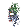
| ||||||||
|---|---|---|---|---|---|---|---|---|---|
| 1 |
| ||||||||
| Unit cell |
|
-Components
| #1: Protein | Mass: 22865.260 Da / Num. of mol.: 2 Source method: isolated from a genetically manipulated source Source: (gene. exp.)  References: UniProt: Q9ZV06, Transferases; Acyltransferases; Transferring groups other than aminoacyl groups #2: Chemical |   Mass: 1533.052 Da / Num. of mol.: 2 / Source method: obtained synthetically / Formula: C42H70N14O32P6S2 / Feature type: SUBJECT OF INVESTIGATION Mass: 1533.052 Da / Num. of mol.: 2 / Source method: obtained synthetically / Formula: C42H70N14O32P6S2 / Feature type: SUBJECT OF INVESTIGATION#3: Water | ChemComp-HOH / |  Mass: 18.015 Da / Num. of mol.: 272 / Source method: isolated from a natural source / Formula: H2O Mass: 18.015 Da / Num. of mol.: 272 / Source method: isolated from a natural source / Formula: H2OHas ligand of interest | Y | |
|---|
-Experimental details
-Experiment
| Experiment | Method: X-RAY DIFFRACTION / Number of used crystals: 1 |
|---|
- Sample preparation
| Crystal | Density Matthews: 2.13 Å3/Da / Density % sol: 42.31 % |
|---|---|
| Crystal grow | Temperature: 291 K / Method: vapor diffusion, sitting drop Details: AtNATA229-226 was concentrated to 10 mg/ml and incubated with a fivefold molar excess of CoA for 18 h on ice. Crystallization drops contained 200 nl protein solution and 200 nl precipitant ...Details: AtNATA229-226 was concentrated to 10 mg/ml and incubated with a fivefold molar excess of CoA for 18 h on ice. Crystallization drops contained 200 nl protein solution and 200 nl precipitant solution. Crystals appeared within 2-18 hours in several conditions. The best diffracting crystals were obtained from the precipitant condition with 0.1 M sodium citrate (pH 5.5), 0.2 M lithium sulfate and 15 % (v/v) ethanol. Crystals were cryo-protected with 20 % glycerol and flash-frozen in liquid nitrogen. |
-Data collection
| Diffraction | Mean temperature: 100 K / Serial crystal experiment: N | |||||||||||||||||||||||||||
|---|---|---|---|---|---|---|---|---|---|---|---|---|---|---|---|---|---|---|---|---|---|---|---|---|---|---|---|---|
| Diffraction source | Source: SYNCHROTRON / Site: ESRF  / Beamline: ID23-1 / Wavelength: 0.972423 Å / Beamline: ID23-1 / Wavelength: 0.972423 Å | |||||||||||||||||||||||||||
| Detector | Type: DECTRIS PILATUS 6M / Detector: PIXEL / Date: Jan 31, 2021 | |||||||||||||||||||||||||||
| Radiation | Protocol: SINGLE WAVELENGTH / Monochromatic (M) / Laue (L): M / Scattering type: x-ray | |||||||||||||||||||||||||||
| Radiation wavelength | Wavelength: 0.972423 Å / Relative weight: 1 | |||||||||||||||||||||||||||
| Reflection | Resolution: 1.45→49.24 Å / Num. obs: 70882 / % possible obs: 99.9 % / Redundancy: 6.1 % / CC1/2: 0.999 / Rmerge(I) obs: 0.067 / Rpim(I) all: 0.029 / Rrim(I) all: 0.073 / Net I/σ(I): 11.5 / Num. measured all: 435296 / Scaling rejects: 96 | |||||||||||||||||||||||||||
| Reflection shell | Diffraction-ID: 1 / Redundancy: 6.2 %
|
- Processing
| Software |
| ||||||||||||||||||||||||||||||||||||||||||||||||||||||||||||||||||||||||||||||||||||||||||||||||||||||||||||||||||||||||||||||||||||||||||||||||||||||||||||||||||||||||||||||||||||||
|---|---|---|---|---|---|---|---|---|---|---|---|---|---|---|---|---|---|---|---|---|---|---|---|---|---|---|---|---|---|---|---|---|---|---|---|---|---|---|---|---|---|---|---|---|---|---|---|---|---|---|---|---|---|---|---|---|---|---|---|---|---|---|---|---|---|---|---|---|---|---|---|---|---|---|---|---|---|---|---|---|---|---|---|---|---|---|---|---|---|---|---|---|---|---|---|---|---|---|---|---|---|---|---|---|---|---|---|---|---|---|---|---|---|---|---|---|---|---|---|---|---|---|---|---|---|---|---|---|---|---|---|---|---|---|---|---|---|---|---|---|---|---|---|---|---|---|---|---|---|---|---|---|---|---|---|---|---|---|---|---|---|---|---|---|---|---|---|---|---|---|---|---|---|---|---|---|---|---|---|---|---|---|---|
| Refinement | Method to determine structure: MOLECULAR REPLACEMENT Starting model: 2fe7 Resolution: 1.45→44.54 Å / SU ML: 0.19 / Cross valid method: THROUGHOUT / σ(F): 1.34 / Phase error: 22.68 / Stereochemistry target values: ML
| ||||||||||||||||||||||||||||||||||||||||||||||||||||||||||||||||||||||||||||||||||||||||||||||||||||||||||||||||||||||||||||||||||||||||||||||||||||||||||||||||||||||||||||||||||||||
| Solvent computation | Shrinkage radii: 0.9 Å / VDW probe radii: 1.11 Å / Solvent model: FLAT BULK SOLVENT MODEL | ||||||||||||||||||||||||||||||||||||||||||||||||||||||||||||||||||||||||||||||||||||||||||||||||||||||||||||||||||||||||||||||||||||||||||||||||||||||||||||||||||||||||||||||||||||||
| Displacement parameters | Biso max: 130.63 Å2 / Biso mean: 37.1394 Å2 / Biso min: 12.34 Å2 | ||||||||||||||||||||||||||||||||||||||||||||||||||||||||||||||||||||||||||||||||||||||||||||||||||||||||||||||||||||||||||||||||||||||||||||||||||||||||||||||||||||||||||||||||||||||
| Refinement step | Cycle: final / Resolution: 1.45→44.54 Å
| ||||||||||||||||||||||||||||||||||||||||||||||||||||||||||||||||||||||||||||||||||||||||||||||||||||||||||||||||||||||||||||||||||||||||||||||||||||||||||||||||||||||||||||||||||||||
| LS refinement shell | Refine-ID: X-RAY DIFFRACTION / Rfactor Rfree error: 0 / Total num. of bins used: 25
|
