 ムービー
ムービー コントローラー


+ データを開く

- 基本情報
基本情報
| 登録情報 | データベース: PDB / ID: 7ovu | |||||||||
|---|---|---|---|---|---|---|---|---|---|---|
| タイトル | Crystal structure of Arabidopsis thaliana NAT9 in complex with AcCoA | |||||||||
 要素 要素 | Acyl-CoA N-acyltransferases (NAT) superfamily protein | |||||||||
 キーワード キーワード | PLANT PROTEIN / GNAT N-acetyltransferase AcCoA PLANT PROTEIN NAT9 | |||||||||
| 機能・相同性 |  機能・相同性情報 機能・相同性情報 | |||||||||
| 生物種 |  | |||||||||
| 手法 |  X線回折 / シンクロトロン / 分子置換 / 解像度: 1.45 Å X線回折 / シンクロトロン / 分子置換 / 解像度: 1.45 Å | |||||||||
 データ登録者 データ登録者 | Layer, D. / Weyer, F.A. / Kopp, J. / Sinning, I. | |||||||||
| 資金援助 |  ドイツ, 2件 ドイツ, 2件
| |||||||||
 引用 引用 | ジャーナル: To Be Published タイトル: Crystal structure of the Arabidopsis thaliana N-acetyltransferase 9 著者: Layer, D. / Weyer, F.A. / Kopp, J. / Sinning, I. | |||||||||
| 履歴 |
|
- 構造の表示
構造の表示
| 構造ビューア | 分子: MolmilJmol/JSmol |
|---|
- ダウンロードとリンク
-ダウンロード
| PDBx/mmCIF形式 | 7ovu.cif.gz | 133.9 KB | 表示 | PDBx/mmCIF形式 |
|---|---|---|---|---|
| PDB形式 | pdb7ovu.ent.gz | 105.3 KB | 表示 | PDB形式 |
| PDBx/mmJSON形式 | 7ovu.json.gz | ツリー表示 | PDBx/mmJSON形式 | |
| その他 |  その他のダウンロード その他のダウンロード |
-検証レポート
| アーカイブディレクトリ | https://data.pdbj.org/pub/pdb/validation_reports/ov/7ovuftp://data.pdbj.org/pub/pdb/validation_reports/ov/7ovu | HTTPS FTP |
|---|
-関連構造データ
| 関連構造データ | 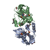 3eo4S S: 精密化の開始モデル |
|---|---|
| 類似構造データ |
-リンク
 PDBj
PDBj


- 集合体
集合体
| 登録構造単位 | 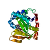
| ||||||||||||
|---|---|---|---|---|---|---|---|---|---|---|---|---|---|
| 1 |
| ||||||||||||
| 単位格子 |
| ||||||||||||
| Components on special symmetry positions |
|
-要素
| #1: タンパク質 | 分子量: 24560.984 Da / 分子数: 1 / 由来タイプ: 組換発現 由来: (組換発現) 遺伝子: At2g04845 / 発現宿主:  |
|---|---|
| #2: 化合物 | ChemComp-ACO /   分子量: 809.571 Da / 分子数: 1 / 由来タイプ: 合成 / 式: C23H38N7O17P3S / タイプ: SUBJECT OF INVESTIGATION 分子量: 809.571 Da / 分子数: 1 / 由来タイプ: 合成 / 式: C23H38N7O17P3S / タイプ: SUBJECT OF INVESTIGATION |
| #3: 水 | ChemComp-HOH /  分子量: 18.015 Da / 分子数: 153 / 由来タイプ: 天然 / 式: H2O 分子量: 18.015 Da / 分子数: 153 / 由来タイプ: 天然 / 式: H2O |
| 研究の焦点であるリガンドがあるか | Y |
-実験情報
-実験
| 実験 | 手法: X線回折 / 使用した結晶の数: 1 |
|---|
- 試料調製
試料調製
| 結晶 | マシュー密度: 2 Å3/Da / 溶媒含有率: 38.53 % |
|---|---|
| 結晶化 | 温度: 291 K / 手法: 蒸気拡散法, シッティングドロップ法 詳細: AtNAT9 was concentrated to 15 mg/ml and incubated with a threefold molar excess of AcCoA for 18 h on ice. Crystallization drops contained 200 nl protein solution and 200 nl precipitant ...詳細: AtNAT9 was concentrated to 15 mg/ml and incubated with a threefold molar excess of AcCoA for 18 h on ice. Crystallization drops contained 200 nl protein solution and 200 nl precipitant solution (0.1 M HEPES (pH 7) and 20 % PEG6000) and crystals appeared after seven days. The crystals were cryo-protected with 20 % glycerol and flash-frozen in liquid nitrogen. |
-データ収集
| 回折 | 平均測定温度: 100 K / Serial crystal experiment: N |
|---|---|
| 放射光源 | 由来: シンクロトロン / サイト: ESRF  / ビームライン: ID29 / 波長: 0.976251 Å / ビームライン: ID29 / 波長: 0.976251 Å |
| 検出器 | タイプ: DECTRIS PILATUS 6M / 検出器: PIXEL / 日付: 2016年12月14日 |
| 放射 | プロトコル: SINGLE WAVELENGTH / 単色(M)・ラウエ(L): M / 散乱光タイプ: x-ray |
| 放射波長 | 波長: 0.976251 Å / 相対比: 1 |
| 反射 | 解像度: 1.45→39.19 Å / Num. obs: 33777 / % possible obs: 99.7 % / 冗長度: 13.1 % / CC1/2: 1 / Rmerge(I) obs: 0.072 / Net I/σ(I): 15.9 |
| 反射 シェル | 解像度: 1.45→1.5 Å / Mean I/σ(I) obs: 1.2 / Num. unique obs: 3306 / CC1/2: 0.509 |
- 解析
解析
| ソフトウェア |
| |||||||||||||||||||||||||||||||||||||||||||||||||||||||||||||||||||||||||||||||||||||||||||
|---|---|---|---|---|---|---|---|---|---|---|---|---|---|---|---|---|---|---|---|---|---|---|---|---|---|---|---|---|---|---|---|---|---|---|---|---|---|---|---|---|---|---|---|---|---|---|---|---|---|---|---|---|---|---|---|---|---|---|---|---|---|---|---|---|---|---|---|---|---|---|---|---|---|---|---|---|---|---|---|---|---|---|---|---|---|---|---|---|---|---|---|---|
| 精密化 | 構造決定の手法: 分子置換 開始モデル: 3eo4 解像度: 1.45→39.19 Å / SU ML: 0.16 / 交差検証法: THROUGHOUT / σ(F): 1.35 / 位相誤差: 20.11 / 立体化学のターゲット値: ML
| |||||||||||||||||||||||||||||||||||||||||||||||||||||||||||||||||||||||||||||||||||||||||||
| 溶媒の処理 | 減衰半径: 0.9 Å / VDWプローブ半径: 1.11 Å / 溶媒モデル: FLAT BULK SOLVENT MODEL | |||||||||||||||||||||||||||||||||||||||||||||||||||||||||||||||||||||||||||||||||||||||||||
| 原子変位パラメータ | Biso max: 93.41 Å2 / Biso mean: 30.7526 Å2 / Biso min: 15.05 Å2 | |||||||||||||||||||||||||||||||||||||||||||||||||||||||||||||||||||||||||||||||||||||||||||
| 精密化ステップ | サイクル: final / 解像度: 1.45→39.19 Å
| |||||||||||||||||||||||||||||||||||||||||||||||||||||||||||||||||||||||||||||||||||||||||||
| LS精密化 シェル | Refine-ID: X-RAY DIFFRACTION / Rfactor Rfree error: 0 / Total num. of bins used: 12
|
