 Movie
Movie Controller


+ Open data

- Basic information
| Entry | Database: PDB / ID: 7otf | |||||||||
|---|---|---|---|---|---|---|---|---|---|---|
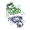
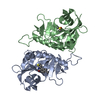
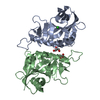
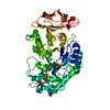
| Title | PARP15 catalytic domain in complex with OUL213 | |||||||||
 Components Components | Protein mono-ADP-ribosyltransferase PARP15 | |||||||||
 Keywords Keywords | TRANSFERASE / Inhibitor / Complex / mono-ADP-ribosyl-transferase | |||||||||
| Function / homology |  Function and homology information Function and homology informationNAD+-protein-aspartate ADP-ribosyltransferase activity / protein poly-ADP-ribosylation / NAD+-protein-glutamate ADP-ribosyltransferase activity / NAD+-protein mono-ADP-ribosyltransferase activity / NAD+ poly-ADP-ribosyltransferase activity / Transferases; Glycosyltransferases; Pentosyltransferases / NAD+ binding / nucleotidyltransferase activity / transcription corepressor activity / negative regulation of gene expression ...NAD+-protein-aspartate ADP-ribosyltransferase activity / protein poly-ADP-ribosylation / NAD+-protein-glutamate ADP-ribosyltransferase activity / NAD+-protein mono-ADP-ribosyltransferase activity / NAD+ poly-ADP-ribosyltransferase activity / Transferases; Glycosyltransferases; Pentosyltransferases / NAD+ binding / nucleotidyltransferase activity / transcription corepressor activity / negative regulation of gene expression / negative regulation of transcription by RNA polymerase II / nucleus / cytoplasm Similarity search - Function | |||||||||
| Biological species |  Homo sapiens (human) Homo sapiens (human) | |||||||||
| Method |  X-RAY DIFFRACTION / SYNCHROTRON / MOLECULAR REPLACEMENT / Resolution: 1.3 Å X-RAY DIFFRACTION / SYNCHROTRON / MOLECULAR REPLACEMENT / Resolution: 1.3 Å | |||||||||
 Authors Authors | Maksimainen, M.M. / Lehtio, L. | |||||||||
| Funding support |  Finland, 2items Finland, 2items
| |||||||||
 Citation Citation | Journal: Bioorg.Med.Chem. / Year: 2021 Title: Analogs of TIQ-A as inhibitors of human mono-ADP-ribosylating PARPs. Authors: Maksimainen, M.M. / Murthy, S. / Sowa, S.T. / Galera-Prat, A. / Rolina, E. / Heiskanen, J.P. / Lehtio, L. | |||||||||
| History |
|
- Structure visualization
| Structure viewer | Molecule: MolmilJmol/JSmol |
|---|
- Downloads & links
-Download
| PDBx/mmCIF format | 7otf.cif.gz | 193.9 KB | Display | PDBx/mmCIF format |
|---|---|---|---|---|
| PDB format | pdb7otf.ent.gz | 153.5 KB | Display | PDB format |
| PDBx/mmJSON format | 7otf.json.gz | Tree view | PDBx/mmJSON format | |
| Others |  Other downloads Other downloads |
-Validation report
| Summary document | 7otf_validation.pdf.gz | 793.1 KB | Display | wwPDB validaton report |
|---|---|---|---|---|
| Full document | 7otf_full_validation.pdf.gz | 796.3 KB | Display | |
| Data in XML | 7otf_validation.xml.gz | 19.4 KB | Display | |
| Data in CIF | 7otf_validation.cif.gz | 29.1 KB | Display | |
| Arichive directory | https://data.pdbj.org/pub/pdb/validation_reports/ot/7otfftp://data.pdbj.org/pub/pdb/validation_reports/ot/7otf | HTTPS FTP |
-Related structure data
| Related structure data |  7oljC  7om1C  7omcC  7oqqC  7ospC  7ossC  7osxC  7othC 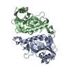 7ouwC  7ouxC 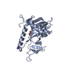 3bljS C: citing same article ( S: Starting model for refinement |
|---|---|
| Similar structure data |


-Links
 PDBj
PDBj- Assembly
| Deposited unit | 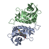
| ||||||||
|---|---|---|---|---|---|---|---|---|---|
| 1 |
| ||||||||
| Unit cell |
|
-Components
| #1: Protein | Mass: 25439.459 Da / Num. of mol.: 2 Source method: isolated from a genetically manipulated source Source: (gene. exp.) Homo sapiens (human) / Gene: PARP15, BAL3 / Production host:  References: UniProt: Q460N3, Transferases; Glycosyltransferases; Pentosyltransferases #2: Chemical | ChemComp-1H9 / |   Mass: 386.262 Da / Num. of mol.: 1 / Source method: obtained synthetically / Formula: C18H12BrNO2S / Feature type: SUBJECT OF INVESTIGATION Mass: 386.262 Da / Num. of mol.: 1 / Source method: obtained synthetically / Formula: C18H12BrNO2S / Feature type: SUBJECT OF INVESTIGATION#3: Chemical | ChemComp-DMS / |   Mass: 78.133 Da / Num. of mol.: 1 / Source method: obtained synthetically / Formula: C2H6OS / Comment: DMSO, precipitant*YM Mass: 78.133 Da / Num. of mol.: 1 / Source method: obtained synthetically / Formula: C2H6OS / Comment: DMSO, precipitant*YM#4: Water | ChemComp-HOH / |  Mass: 18.015 Da / Num. of mol.: 334 / Source method: isolated from a natural source / Formula: H2O Mass: 18.015 Da / Num. of mol.: 334 / Source method: isolated from a natural source / Formula: H2OHas ligand of interest | Y | |
|---|
-Experimental details
-Experiment
| Experiment | Method: X-RAY DIFFRACTION / Number of used crystals: 1 |
|---|
- Sample preparation
| Crystal | Density Matthews: 2.42 Å3/Da / Density % sol: 49.26 % |
|---|---|
| Crystal grow | Temperature: 295 K / Method: vapor diffusion, sitting drop / pH: 7.5 / Details: 0.2 M ammonium acetate, 20% (w/v) PEG 3350 |
-Data collection
| Diffraction | Mean temperature: 100 K / Serial crystal experiment: N |
|---|---|
| Diffraction source | Source: SYNCHROTRON / Site: Diamond  / Beamline: I03 / Wavelength: 0.97625 Å / Beamline: I03 / Wavelength: 0.97625 Å |
| Detector | Type: DECTRIS EIGER2 X 16M / Detector: PIXEL / Date: Aug 6, 2019 |
| Radiation | Protocol: SINGLE WAVELENGTH / Monochromatic (M) / Laue (L): M / Scattering type: x-ray |
| Radiation wavelength | Wavelength: 0.97625 Å / Relative weight: 1 |
| Reflection | Resolution: 1.3→50 Å / Num. obs: 122067 / % possible obs: 99.7 % / Redundancy: 13 % / CC1/2: 1 / Net I/σ(I): 23.24 |
| Reflection shell | Resolution: 1.3→1.33 Å / Num. unique obs: 8626 / CC1/2: 0.72 |
- Processing
| Software |
| |||||||||||||||||||||||||||||||||||||||||||||||||||||||||||||||||
|---|---|---|---|---|---|---|---|---|---|---|---|---|---|---|---|---|---|---|---|---|---|---|---|---|---|---|---|---|---|---|---|---|---|---|---|---|---|---|---|---|---|---|---|---|---|---|---|---|---|---|---|---|---|---|---|---|---|---|---|---|---|---|---|---|---|---|
| Refinement | Method to determine structure: MOLECULAR REPLACEMENT Starting model: 3BLJ Resolution: 1.3→43.61 Å / Cor.coef. Fo:Fc: 0.977 / Cor.coef. Fo:Fc free: 0.972 / SU B: 1.887 / SU ML: 0.034 / Cross valid method: THROUGHOUT / σ(F): 0 / ESU R: 0.041 / ESU R Free: 0.041 / Stereochemistry target values: MAXIMUM LIKELIHOOD Details: HYDROGENS HAVE BEEN ADDED IN THE RIDING POSITIONS U VALUES : REFINED INDIVIDUALLY
| |||||||||||||||||||||||||||||||||||||||||||||||||||||||||||||||||
| Solvent computation | Ion probe radii: 0.8 Å / Shrinkage radii: 0.8 Å / VDW probe radii: 1.2 Å / Solvent model: MASK | |||||||||||||||||||||||||||||||||||||||||||||||||||||||||||||||||
| Displacement parameters | Biso max: 90.82 Å2 / Biso mean: 22.038 Å2 / Biso min: 11.83 Å2
| |||||||||||||||||||||||||||||||||||||||||||||||||||||||||||||||||
| Refinement step | Cycle: final / Resolution: 1.3→43.61 Å
| |||||||||||||||||||||||||||||||||||||||||||||||||||||||||||||||||
| Refine LS restraints |
| |||||||||||||||||||||||||||||||||||||||||||||||||||||||||||||||||
| LS refinement shell | Resolution: 1.3→1.334 Å / Rfactor Rfree error: 0 / Total num. of bins used: 20
|
