 Movie
Movie Controller


+ Open data

- Basic information
| Entry | Database: PDB / ID: 7oqr | ||||||
|---|---|---|---|---|---|---|---|
| Title | Crystal structure of Trypanosoma cruzi peroxidase | ||||||
 Components Components | Ascorbate peroxidase | ||||||
 Keywords Keywords | OXIDOREDUCTASE / heme peroxidase | ||||||
| Function / homology |  Function and homology information Function and homology informationL-ascorbate peroxidase activity / cytochrome-c peroxidase / response to reactive oxygen species / hydrogen peroxide catabolic process / mitochondrial intermembrane space / cellular response to oxidative stress / mitochondrial matrix / heme binding / metal ion binding Similarity search - Function | ||||||
| Biological species |  | ||||||
| Method |  X-RAY DIFFRACTION / SYNCHROTRON / MOLECULAR REPLACEMENT / Resolution: 1.76 Å X-RAY DIFFRACTION / SYNCHROTRON / MOLECULAR REPLACEMENT / Resolution: 1.76 Å | ||||||
 Authors Authors | Freeman, S.L. / Kwon, H. / Skafar, V. / Fielding, A.J. / Martinez, A. / Piacenza, L. / Radi, R. / Raven, E.L. | ||||||
| Funding support |  United Kingdom, 1items United Kingdom, 1items
| ||||||
 Citation Citation | Journal: J.Biol.Chem. / Year: 2022 Title: Crystal structure of Trypanosoma cruzi heme peroxidase and characterization of its substrate specificity and compound I intermediate. Authors: Freeman, S.L. / Skafar, V. / Kwon, H. / Fielding, A.J. / Moody, P.C.E. / Martinez, A. / Issoglio, F.M. / Inchausti, L. / Smircich, P. / Zeida, A. / Piacenza, L. / Radi, R. / Raven, E.L. | ||||||
| History |
|
- Structure visualization
| Structure viewer | Molecule: MolmilJmol/JSmol |
|---|
- Downloads & links
-Download
| PDBx/mmCIF format | 7oqr.cif.gz | 139 KB | Display | PDBx/mmCIF format |
|---|---|---|---|---|
| PDB format | pdb7oqr.ent.gz | 106.7 KB | Display | PDB format |
| PDBx/mmJSON format | 7oqr.json.gz | Tree view | PDBx/mmJSON format | |
| Others |  Other downloads Other downloads |
-Validation report
| Summary document | 7oqr_validation.pdf.gz | 1.4 MB | Display | wwPDB validaton report |
|---|---|---|---|---|
| Full document | 7oqr_full_validation.pdf.gz | 1.4 MB | Display | |
| Data in XML | 7oqr_validation.xml.gz | 27.4 KB | Display | |
| Data in CIF | 7oqr_validation.cif.gz | 40 KB | Display | |
| Arichive directory | https://data.pdbj.org/pub/pdb/validation_reports/oq/7oqrftp://data.pdbj.org/pub/pdb/validation_reports/oq/7oqr | HTTPS FTP |
-Related structure data
| Related structure data | 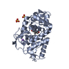 7optSC S: Starting model for refinement C: citing same article ( |
|---|---|
| Similar structure data |
-Links
 PDBj
PDBj


- Assembly
| Deposited unit | 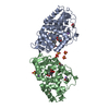
| ||||||||
|---|---|---|---|---|---|---|---|---|---|
| 1 | 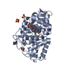
| ||||||||
| 2 | 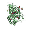
| ||||||||
| Unit cell |
|
-Components
-Protein , 1 types, 2 molecules AB
| #1: Protein | Mass: 36511.316 Da / Num. of mol.: 2 Source method: isolated from a genetically manipulated source Source: (gene. exp.)  |
|---|
-Non-polymers , 7 types, 442 molecules 


| #2: Chemical |  Mass: 616.487 Da / Num. of mol.: 2 / Source method: obtained synthetically / Formula: C34H32FeN4O4 / Feature type: SUBJECT OF INVESTIGATION Mass: 616.487 Da / Num. of mol.: 2 / Source method: obtained synthetically / Formula: C34H32FeN4O4 / Feature type: SUBJECT OF INVESTIGATION#3: Chemical | ChemComp-SO4 /  Mass: 96.063 Da / Num. of mol.: 5 / Source method: obtained synthetically / Formula: SO4 Mass: 96.063 Da / Num. of mol.: 5 / Source method: obtained synthetically / Formula: SO4#4: Chemical | ChemComp-GOL /  Mass: 92.094 Da / Num. of mol.: 5 / Source method: obtained synthetically / Formula: C3H8O3 Mass: 92.094 Da / Num. of mol.: 5 / Source method: obtained synthetically / Formula: C3H8O3#5: Chemical | ChemComp-NA /  Mass: 22.990 Da / Num. of mol.: 4 / Source method: obtained synthetically / Formula: Na Mass: 22.990 Da / Num. of mol.: 4 / Source method: obtained synthetically / Formula: Na#6: Chemical | ChemComp-OXY / |  Mass: 31.999 Da / Num. of mol.: 1 / Source method: obtained synthetically / Formula: O2 Mass: 31.999 Da / Num. of mol.: 1 / Source method: obtained synthetically / Formula: O2#7: Chemical | ChemComp-ACT / |  Mass: 59.044 Da / Num. of mol.: 1 / Source method: obtained synthetically / Formula: C2H3O2 Mass: 59.044 Da / Num. of mol.: 1 / Source method: obtained synthetically / Formula: C2H3O2#8: Water | ChemComp-HOH / | Mass: 18.015 Da / Num. of mol.: 424 / Source method: isolated from a natural source / Formula: H2O |
|---|
-Details
| Has ligand of interest | Y |
|---|
-Experimental details
-Experiment
| Experiment | Method: X-RAY DIFFRACTION / Number of used crystals: 1 |
|---|
- Sample preparation
| Crystal | Density Matthews: 2.58 Å3/Da / Density % sol: 52.31 % |
|---|---|
| Crystal grow | Temperature: 297 K / Method: vapor diffusion, hanging drop / Details: ammonium sulfate, sodium acetate |
-Data collection
| Diffraction | Mean temperature: 100 K / Serial crystal experiment: N | |||||||||||||||
|---|---|---|---|---|---|---|---|---|---|---|---|---|---|---|---|---|
| Diffraction source | Source: SYNCHROTRON / Site: Diamond / Beamline: I04 / Wavelength: 0.9795 Å | |||||||||||||||
| Detector | Type: DECTRIS EIGER2 X 16M / Detector: PIXEL / Date: Dec 5, 2019 | |||||||||||||||
| Radiation | Protocol: SINGLE WAVELENGTH / Monochromatic (M) / Laue (L): M / Scattering type: x-ray | |||||||||||||||
| Radiation wavelength | Wavelength: 0.9795 Å / Relative weight: 1 | |||||||||||||||
| Reflection twin |
| |||||||||||||||
| Reflection | Resolution: 1.76→29.28 Å / Num. obs: 76005 / % possible obs: 99.8 % / Redundancy: 20 % / Rmerge(I) obs: 0.086 / Net I/σ(I): 22.5 | |||||||||||||||
| Reflection shell | Resolution: 1.76→1.8 Å / Rmerge(I) obs: 0.785 / Num. unique obs: 4170 |
- Processing
| Software |
| |||||||||||||||||||||||||||||||||||||||||||||||||||||||||||||||||
|---|---|---|---|---|---|---|---|---|---|---|---|---|---|---|---|---|---|---|---|---|---|---|---|---|---|---|---|---|---|---|---|---|---|---|---|---|---|---|---|---|---|---|---|---|---|---|---|---|---|---|---|---|---|---|---|---|---|---|---|---|---|---|---|---|---|---|
| Refinement | Method to determine structure: MOLECULAR REPLACEMENT Starting model: 7OPT Resolution: 1.76→29.28 Å / Cor.coef. Fo:Fc: 0.98 / Cor.coef. Fo:Fc free: 0.968 / SU B: 1.274 / SU ML: 0.041 / Cross valid method: THROUGHOUT / σ(F): 0 / ESU R: 0.014 / ESU R Free: 0.016 / Stereochemistry target values: MAXIMUM LIKELIHOOD
| |||||||||||||||||||||||||||||||||||||||||||||||||||||||||||||||||
| Solvent computation | Ion probe radii: 0.8 Å / Shrinkage radii: 0.8 Å / VDW probe radii: 1.2 Å / Solvent model: MASK | |||||||||||||||||||||||||||||||||||||||||||||||||||||||||||||||||
| Displacement parameters | Biso max: 81.36 Å2 / Biso mean: 25.733 Å2 / Biso min: 15.17 Å2
| |||||||||||||||||||||||||||||||||||||||||||||||||||||||||||||||||
| Refinement step | Cycle: final / Resolution: 1.76→29.28 Å
| |||||||||||||||||||||||||||||||||||||||||||||||||||||||||||||||||
| Refine LS restraints |
| |||||||||||||||||||||||||||||||||||||||||||||||||||||||||||||||||
| LS refinement shell | Resolution: 1.762→1.808 Å / Rfactor Rfree error: 0 / Total num. of bins used: 20
|
