 Movie
Movie Controller


+ Open data

- Basic information
| Entry | Database: PDB / ID: 7ol2 | ||||||
|---|---|---|---|---|---|---|---|
| Title | Crystal structure of mouse contactin 1 immunoglobulin domains | ||||||
 Components Components | Contactin-1 | ||||||
 Keywords Keywords | CELL ADHESION / Zipper / Dimer / Glycoprotein / Immunoglobulin cell adhesion protein Neural cell adhesion protein / Horseshoe | ||||||
| Function / homology |  Function and homology information Function and homology informationcentral nervous system myelin formation / positive regulation of sodium ion transport / side of membrane / Notch signaling pathway / myelination / cerebellum development / locomotory behavior / positive regulation of neuron projection development / neuron projection development / myelin sheath ...central nervous system myelin formation / positive regulation of sodium ion transport / side of membrane / Notch signaling pathway / myelination / cerebellum development / locomotory behavior / positive regulation of neuron projection development / neuron projection development / myelin sheath / carbohydrate binding / presynaptic membrane / gene expression / postsynaptic membrane / cell adhesion / synapse / positive regulation of gene expression / glutamatergic synapse / plasma membrane Similarity search - Function | ||||||
| Biological species |  | ||||||
| Method |  X-RAY DIFFRACTION / SYNCHROTRON / MOLECULAR REPLACEMENT / Resolution: 3.89 Å X-RAY DIFFRACTION / SYNCHROTRON / MOLECULAR REPLACEMENT / Resolution: 3.89 Å | ||||||
 Authors Authors | Chataigner, L.M.P. / Janssen, B.J.C. | ||||||
| Funding support |  Netherlands, 1items Netherlands, 1items
| ||||||
 Citation Citation | Journal: Nat Commun / Year: 2022 Title: Structural insights into the contactin 1 - neurofascin 155 adhesion complex. Authors: Chataigner, L.M.P. / Gogou, C. / den Boer, M.A. / Frias, C.P. / Thies-Weesie, D.M.E. / Granneman, J.C.M. / Heck, A.J.R. / Meijer, D.H. / Janssen, B.J.C. | ||||||
| History |
|
- Structure visualization
| Structure viewer | Molecule: MolmilJmol/JSmol |
|---|
- Downloads & links
-Download
| PDBx/mmCIF format | 7ol2.cif.gz | 248.4 KB | Display | PDBx/mmCIF format |
|---|---|---|---|---|
| PDB format | pdb7ol2.ent.gz | 197.5 KB | Display | PDB format |
| PDBx/mmJSON format | 7ol2.json.gz | Tree view | PDBx/mmJSON format | |
| Others |  Other downloads Other downloads |
-Validation report
| Arichive directory | https://data.pdbj.org/pub/pdb/validation_reports/ol/7ol2ftp://data.pdbj.org/pub/pdb/validation_reports/ol/7ol2 | HTTPS FTP |
|---|
-Related structure data
| Related structure data | 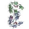 7ok5C 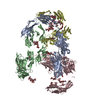 7ol4C 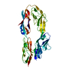 2om5S S: Starting model for refinement C: citing same article ( |
|---|---|
| Similar structure data |
-Links
 PDBj
PDBj


- Assembly
| Deposited unit | 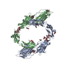
| |||||||||||||||||||||||||||||||||||||||||||||||||||
|---|---|---|---|---|---|---|---|---|---|---|---|---|---|---|---|---|---|---|---|---|---|---|---|---|---|---|---|---|---|---|---|---|---|---|---|---|---|---|---|---|---|---|---|---|---|---|---|---|---|---|---|---|
| 1 |
| |||||||||||||||||||||||||||||||||||||||||||||||||||
| Unit cell |
| |||||||||||||||||||||||||||||||||||||||||||||||||||
| Noncrystallographic symmetry (NCS) | NCS domain:
NCS domain segments:
|
-Components
| #1: Protein | Mass: 66907.609 Da / Num. of mol.: 2 Source method: isolated from a genetically manipulated source Source: (gene. exp.)  Homo sapiens (human) / References: UniProt: P12960 Homo sapiens (human) / References: UniProt: P12960#2: Polysaccharide | Source method: isolated from a genetically manipulated source #3: Polysaccharide | Source method: isolated from a genetically manipulated source #4: Polysaccharide | Source method: isolated from a genetically manipulated source #5: Sugar | ChemComp-NAG /   Type: D-saccharide, beta linking / Mass: 221.208 Da / Num. of mol.: 5 / Source method: obtained synthetically / Formula: C8H15NO6 / Feature type: SUBJECT OF INVESTIGATION Type: D-saccharide, beta linking / Mass: 221.208 Da / Num. of mol.: 5 / Source method: obtained synthetically / Formula: C8H15NO6 / Feature type: SUBJECT OF INVESTIGATIONHas ligand of interest | Y | Has protein modification | Y | |
|---|
-Experimental details
-Experiment
| Experiment | Method: X-RAY DIFFRACTION / Number of used crystals: 1 |
|---|
- Sample preparation
| Crystal | Density Matthews: 4.23 Å3/Da / Density % sol: 70.95 % |
|---|---|
| Crystal grow | Temperature: 277.15 K / Method: vapor diffusion, sitting drop / pH: 5.9 Details: 20% w/v PEG 3350 and 0.2 M Magnesium nitrate hexahydrate pH 5.9 |
-Data collection
| Diffraction | Mean temperature: 100 K / Serial crystal experiment: N |
|---|---|
| Diffraction source | Source: SYNCHROTRON / Site: Diamond  / Beamline: I24 / Wavelength: 0.9686 Å / Beamline: I24 / Wavelength: 0.9686 Å |
| Detector | Type: DECTRIS PILATUS3 6M / Detector: PIXEL / Date: Jan 31, 2019 |
| Radiation | Protocol: SINGLE WAVELENGTH / Monochromatic (M) / Laue (L): M / Scattering type: x-ray |
| Radiation wavelength | Wavelength: 0.9686 Å / Relative weight: 1 |
| Reflection | Resolution: 3.89→108.09 Å / Num. obs: 20221 / % possible obs: 93.9 % / Redundancy: 9.3 % / CC1/2: 0.961 / Net I/σ(I): 4.4 |
| Reflection shell | Resolution: 3.89→4.27 Å / Mean I/σ(I) obs: 1.5 / Num. unique obs: 3909 / CC1/2: 0.524 |
- Processing
| Software |
| ||||||||||||||||||||||||||||||||||||||||||||||||||||||||
|---|---|---|---|---|---|---|---|---|---|---|---|---|---|---|---|---|---|---|---|---|---|---|---|---|---|---|---|---|---|---|---|---|---|---|---|---|---|---|---|---|---|---|---|---|---|---|---|---|---|---|---|---|---|---|---|---|---|
| Refinement | Method to determine structure: MOLECULAR REPLACEMENT Starting model: 2OM5 Resolution: 3.89→107.94 Å / SU ML: 0.39 / Cross valid method: THROUGHOUT / σ(F): 1.34 / Phase error: 26.3 / Stereochemistry target values: ML
| ||||||||||||||||||||||||||||||||||||||||||||||||||||||||
| Solvent computation | Shrinkage radii: 0.9 Å / VDW probe radii: 1.11 Å / Solvent model: FLAT BULK SOLVENT MODEL | ||||||||||||||||||||||||||||||||||||||||||||||||||||||||
| Displacement parameters | Biso max: 201.77 Å2 / Biso mean: 105.278 Å2 / Biso min: 39.79 Å2 | ||||||||||||||||||||||||||||||||||||||||||||||||||||||||
| Refinement step | Cycle: final / Resolution: 3.89→107.94 Å
| ||||||||||||||||||||||||||||||||||||||||||||||||||||||||
| Refine LS restraints NCS |
| ||||||||||||||||||||||||||||||||||||||||||||||||||||||||
| LS refinement shell | Refine-ID: X-RAY DIFFRACTION / Rfactor Rfree error: 0 / Total num. of bins used: 7
|
