 Movie
Movie Controller


+ Open data

- Basic information
| Entry | Database: PDB / ID: 7og8 | ||||||
|---|---|---|---|---|---|---|---|
| Title | Wild-type Hfq protein from Neisseria meningitidis | ||||||
 Components Components | RNA-binding protein Hfq | ||||||
 Keywords Keywords | RNA BINDING PROTEIN / sRNA / mRNA / annealing | ||||||
| Function / homology | RNA-binding protein Hfq / Hfq protein / : / Sm domain profile. / LSM domain superfamily / regulation of DNA-templated transcription / RNA binding / RNA-binding protein Hfq Function and homology information Function and homology information | ||||||
| Biological species | Neisseria meningitidis serogroup C / serotype 2a | ||||||
| Method |  X-RAY DIFFRACTION / SYNCHROTRON / MOLECULAR REPLACEMENT / Resolution: 1.4 Å X-RAY DIFFRACTION / SYNCHROTRON / MOLECULAR REPLACEMENT / Resolution: 1.4 Å | ||||||
 Authors Authors | Moche, M. / Karlsson, J. / Loh, E. | ||||||
 Citation Citation | Journal: To Be Published Title: Crystal structures of wild type, Q9A and R17A single mutant Hfq structures from Neisseria meningitidis Authors: Karlsson, J. / Moche, M. / Loh, E. | ||||||
| History |
|
- Structure visualization
| Structure viewer | Molecule: MolmilJmol/JSmol |
|---|
- Downloads & links
-Download
| PDBx/mmCIF format | 7og8.cif.gz | 28.5 KB | Display | PDBx/mmCIF format |
|---|---|---|---|---|
| PDB format | pdb7og8.ent.gz | 17.3 KB | Display | PDB format |
| PDBx/mmJSON format | 7og8.json.gz | Tree view | PDBx/mmJSON format | |
| Others |  Other downloads Other downloads |
-Validation report
| Arichive directory | https://data.pdbj.org/pub/pdb/validation_reports/og/7og8ftp://data.pdbj.org/pub/pdb/validation_reports/og/7og8 | HTTPS FTP |
|---|
-Related structure data
| Related structure data | 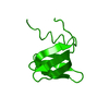 7ogwC 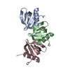 7oh8C 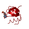 4pnoS S: Starting model for refinement C: citing same article ( |
|---|---|
| Similar structure data |
-Links
 PDBj
PDBj

- Assembly
| Deposited unit | 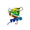
| ||||||||||||
|---|---|---|---|---|---|---|---|---|---|---|---|---|---|
| 1 | x 6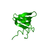
| ||||||||||||
| Unit cell |
| ||||||||||||
| Components on special symmetry positions |
|
-Components
| #1: Protein | Mass: 7676.911 Da / Num. of mol.: 1 Source method: isolated from a genetically manipulated source Source: (gene. exp.)  Neisseria meningitidis serogroup C / serotype 2a (strain ATCC 700532 / DSM 15464 / FAM18) (bacteria) Neisseria meningitidis serogroup C / serotype 2a (strain ATCC 700532 / DSM 15464 / FAM18) (bacteria)Strain: ATCC 700532 / DSM 15464 / FAM18 / Gene: hfq, NMC0702 / Production host: |
|---|---|
| #2: Water | ChemComp-HOH /  Mass: 18.015 Da / Num. of mol.: 53 / Source method: isolated from a natural source / Formula: H2O Mass: 18.015 Da / Num. of mol.: 53 / Source method: isolated from a natural source / Formula: H2O |
-Experimental details
-Experiment
| Experiment | Method: X-RAY DIFFRACTION / Number of used crystals: 1 |
|---|
- Sample preparation
| Crystal | Density Matthews: 1.89 Å3/Da / Density % sol: 35.03 % |
|---|---|
| Crystal grow | Temperature: 293 K / Method: vapor diffusion, sitting drop / pH: 6.1 Details: PEG3350, sodium nitrate, glycerol, bis-tris propane PH range: 6.1-7.4 |
-Data collection
| Diffraction | Mean temperature: 100 K / Serial crystal experiment: N | ||||||||||||||||||||||||||||||
|---|---|---|---|---|---|---|---|---|---|---|---|---|---|---|---|---|---|---|---|---|---|---|---|---|---|---|---|---|---|---|---|
| Diffraction source | Source: SYNCHROTRON / Site: BESSY  / Beamline: 14.2 / Wavelength: 0.9184 Å / Beamline: 14.2 / Wavelength: 0.9184 Å | ||||||||||||||||||||||||||||||
| Detector | Type: DECTRIS PILATUS3 2M / Detector: PIXEL / Date: Oct 19, 2017 | ||||||||||||||||||||||||||||||
| Radiation | Protocol: SINGLE WAVELENGTH / Monochromatic (M) / Laue (L): M / Scattering type: x-ray | ||||||||||||||||||||||||||||||
| Radiation wavelength | Wavelength: 0.9184 Å / Relative weight: 1 | ||||||||||||||||||||||||||||||
| Reflection | Resolution: 1.4→30.27 Å / Num. obs: 11543 / % possible obs: 100 % / Redundancy: 18.3 % / CC1/2: 0.999 / Rmerge(I) obs: 0.113 / Rpim(I) all: 0.027 / Rrim(I) all: 0.116 / Net I/σ(I): 13.7 / Num. measured all: 211386 / Scaling rejects: 1406 | ||||||||||||||||||||||||||||||
| Reflection shell | Diffraction-ID: 1
|
- Processing
| Software |
| ||||||||||||||||||||||||||||||||||||||||||||||||||||||||||||
|---|---|---|---|---|---|---|---|---|---|---|---|---|---|---|---|---|---|---|---|---|---|---|---|---|---|---|---|---|---|---|---|---|---|---|---|---|---|---|---|---|---|---|---|---|---|---|---|---|---|---|---|---|---|---|---|---|---|---|---|---|---|
| Refinement | Method to determine structure: MOLECULAR REPLACEMENT Starting model: 4PNO Resolution: 1.4→20.35 Å / Cor.coef. Fo:Fc: 0.922 / Cor.coef. Fo:Fc free: 0.894 / SU B: 1.56 / SU ML: 0.064 / Cross valid method: THROUGHOUT / σ(F): 0 / ESU R: 0.093 / ESU R Free: 0.093 / Stereochemistry target values: MAXIMUM LIKELIHOOD Details: HYDROGENS HAVE BEEN ADDED IN THE RIDING POSITIONS U VALUES : REFINED INDIVIDUALLY
| ||||||||||||||||||||||||||||||||||||||||||||||||||||||||||||
| Solvent computation | Ion probe radii: 0.8 Å / Shrinkage radii: 0.8 Å / VDW probe radii: 1.2 Å / Solvent model: MASK | ||||||||||||||||||||||||||||||||||||||||||||||||||||||||||||
| Displacement parameters | Biso max: 56.97 Å2 / Biso mean: 14.642 Å2 / Biso min: 6.66 Å2
| ||||||||||||||||||||||||||||||||||||||||||||||||||||||||||||
| Refinement step | Cycle: final / Resolution: 1.4→20.35 Å
| ||||||||||||||||||||||||||||||||||||||||||||||||||||||||||||
| Refine LS restraints |
| ||||||||||||||||||||||||||||||||||||||||||||||||||||||||||||
| LS refinement shell | Resolution: 1.4→1.436 Å / Rfactor Rfree error: 0 / Total num. of bins used: 20
|
