 Movie
Movie Controller

[English] 日本語
 Yorodumi
- PDB-7ocj: Crystal structure of E.coli LexA in complex with nanobody NbSOS2(... -
+ Open data

- Basic information
| Entry | Database: PDB / ID: 7ocj | ||||||
|---|---|---|---|---|---|---|---|
| Title | Crystal structure of E.coli LexA in complex with nanobody NbSOS2(Nb14509) | ||||||
 Components Components |
| ||||||
 Keywords Keywords | TRANSCRIPTION / Transcriptional repressor DNA binding autoproteolysis Nanobodies | ||||||
| Function / homology |  Function and homology information Function and homology informationrepressor LexA / SOS response / DNA replication / serine-type endopeptidase activity / DNA repair / negative regulation of DNA-templated transcription / proteolysis / DNA binding Similarity search - Function | ||||||
| Biological species |   | ||||||
| Method |  X-RAY DIFFRACTION / SYNCHROTRON / MOLECULAR REPLACEMENT / Resolution: 2.7 Å X-RAY DIFFRACTION / SYNCHROTRON / MOLECULAR REPLACEMENT / Resolution: 2.7 Å | ||||||
 Authors Authors | Maso, L. / Vascon, F. / Chinellato, M. / Pardon, E. / Steyaert, J. / Angelini, A. / Tondi, D. / Cendron, L. | ||||||
| Funding support |  Italy, 1items Italy, 1items
| ||||||
 Citation Citation | Journal: Structure / Year: 2022 Title: Nanobodies targeting LexA autocleavage disclose a novel suppression strategy of SOS-response pathway. Authors: Maso, L. / Vascon, F. / Chinellato, M. / Goormaghtigh, F. / Bellio, P. / Campagnaro, E. / Van Melderen, L. / Ruzzene, M. / Pardon, E. / Angelini, A. / Celenza, G. / Steyaert, J. / Tondi, D. / Cendron, L. | ||||||
| History |
|
- Structure visualization
| Structure viewer | Molecule: MolmilJmol/JSmol |
|---|
- Downloads & links
-Download
| PDBx/mmCIF format | 7ocj.cif.gz | 376.6 KB | Display | PDBx/mmCIF format |
|---|---|---|---|---|
| PDB format | pdb7ocj.ent.gz | Display | PDB format | |
| PDBx/mmJSON format | 7ocj.json.gz | Tree view | PDBx/mmJSON format | |
| Others |  Other downloads Other downloads |
-Validation report
| Arichive directory | https://data.pdbj.org/pub/pdb/validation_reports/oc/7ocjftp://data.pdbj.org/pub/pdb/validation_reports/oc/7ocj | HTTPS FTP |
|---|
-Related structure data
| Related structure data | 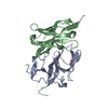 7b5gC  7zraC  1jhfS  7bg2 C: citing same article ( S: Starting model for refinement |
|---|---|
| Similar structure data |
-Links
 PDBj
PDBj

- Assembly
| Deposited unit | 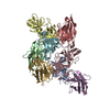
| ||||||||
|---|---|---|---|---|---|---|---|---|---|
| 1 | 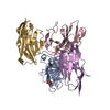
| ||||||||
| 2 | 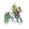
| ||||||||
| Unit cell |
|
-Components
| #1: Antibody | Mass: 12422.886 Da / Num. of mol.: 4 Source method: isolated from a genetically manipulated source Source: (gene. exp.) #2: Protein | Mass: 24560.086 Da / Num. of mol.: 4 Source method: isolated from a genetically manipulated source Details: missing residues are not visible in the electron density maps Source: (gene. exp.) #3: Chemical | ChemComp-EDO / |   Mass: 62.068 Da / Num. of mol.: 1 / Source method: obtained synthetically / Formula: C2H6O2 Mass: 62.068 Da / Num. of mol.: 1 / Source method: obtained synthetically / Formula: C2H6O2#4: Water | ChemComp-HOH / |  Mass: 18.015 Da / Num. of mol.: 46 / Source method: isolated from a natural source / Formula: H2O Mass: 18.015 Da / Num. of mol.: 46 / Source method: isolated from a natural source / Formula: H2OHas ligand of interest | N | Has protein modification | Y | |
|---|
-Experimental details
-Experiment
| Experiment | Method: X-RAY DIFFRACTION / Number of used crystals: 1 |
|---|
- Sample preparation
| Crystal | Density Matthews: 2.47 Å3/Da / Density % sol: 50.27 % |
|---|---|
| Crystal grow | Temperature: 293 K / Method: vapor diffusion, sitting drop Details: 0.2 M lithium sulfate, 0.1 M sodium acetate pH 4.5, 30 % w/v PEG8000 |
-Data collection
| Diffraction | Mean temperature: 100 K / Serial crystal experiment: N |
|---|---|
| Diffraction source | Source: SYNCHROTRON / Site: ESRF  / Beamline: ID30B / Wavelength: 0.96546 Å / Beamline: ID30B / Wavelength: 0.96546 Å |
| Detector | Type: DECTRIS PILATUS3 2M / Detector: PIXEL / Date: Apr 14, 2021 |
| Radiation | Protocol: SINGLE WAVELENGTH / Monochromatic (M) / Laue (L): M / Scattering type: x-ray |
| Radiation wavelength | Wavelength: 0.96546 Å / Relative weight: 1 |
| Reflection | Resolution: 2.7→48.79 Å / Num. obs: 28568 / % possible obs: 96.2 % / Redundancy: 7.4 % / CC1/2: 0.995 / Rmerge(I) obs: 0.126 / Net I/σ(I): 11.1 |
| Reflection shell | Resolution: 2.7→2.83 Å / Rmerge(I) obs: 0.73 / Mean I/σ(I) obs: 1.2 / Num. unique obs: 8056 / CC1/2: 0.558 |
- Processing
| Software |
| ||||||||||||||||||
|---|---|---|---|---|---|---|---|---|---|---|---|---|---|---|---|---|---|---|---|
| Refinement | Method to determine structure: MOLECULAR REPLACEMENT Starting model: 1JHF Resolution: 2.7→48.25 Å / Cross valid method: THROUGHOUT
| ||||||||||||||||||
| Displacement parameters | Biso max: 93.72 Å2 / Biso mean: 47.6129 Å2 / Biso min: 30 Å2 | ||||||||||||||||||
| Refinement step | Cycle: LAST / Resolution: 2.7→48.25 Å
|
