 Movie
Movie Controller

[English] 日本語
 Yorodumi
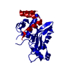
- PDB-7ob6: CPR-C4 - a conserved novel protease from the Candidate Phyla Radiation -
+ Open data

- Basic information
| Entry | Database: PDB / ID: 7ob6 | ||||||
|---|---|---|---|---|---|---|---|
| Title | CPR-C4 - a conserved novel protease from the Candidate Phyla Radiation | ||||||
 Components Components | CPR-C4 | ||||||
 Keywords Keywords | HYDROLASE / Protease / cysteine protease / Zn | ||||||
| Biological species |  candidate division CPR1 (bacteria) candidate division CPR1 (bacteria) | ||||||
| Method |  X-RAY DIFFRACTION / SYNCHROTRON / MAD / Resolution: 2.598 Å X-RAY DIFFRACTION / SYNCHROTRON / MAD / Resolution: 2.598 Å | ||||||
 Authors Authors | Cornish, K.A.S. / Pohl, E. | ||||||
| Funding support | European Union, 1items
| ||||||
 Citation Citation | Journal: J.Biol.Chem. / Year: 2022 Title: CPR-C4 is a highly conserved novel protease from the Candidate Phyla Radiation with remote structural homology to human vasohibins. Authors: Cornish, K.A.S. / Lange, J. / Aevarsson, A. / Pohl, E. | ||||||
| History |
|
- Structure visualization
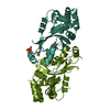
| Structure viewer | Molecule:  MolmilJmol/JSmol MolmilJmol/JSmol |
|---|
- Downloads & links
-Download
| PDBx/mmCIF format | 7ob6.cif.gz | 104.3 KB | Display | PDBx/mmCIF format |
|---|---|---|---|---|
| PDB format | pdb7ob6.ent.gz | 74.8 KB | Display | PDB format |
| PDBx/mmJSON format | 7ob6.json.gz | Tree view | PDBx/mmJSON format | |
| Others |  Other downloads Other downloads |
-Validation report
| Summary document | 7ob6_validation.pdf.gz | 1.1 MB | Display | wwPDB validaton report |
|---|---|---|---|---|
| Full document | 7ob6_full_validation.pdf.gz | 1.1 MB | Display | |
| Data in XML | 7ob6_validation.xml.gz | 17.3 KB | Display | |
| Data in CIF | 7ob6_validation.cif.gz | 23.6 KB | Display | |
| Arichive directory | https://data.pdbj.org/pub/pdb/validation_reports/ob/7ob6ftp://data.pdbj.org/pub/pdb/validation_reports/ob/7ob6 | HTTPS FTP |
-Related structure data
-Links
 PDBj
PDBj- Assembly
| Deposited unit | 
| |||||||||||||||||||||
|---|---|---|---|---|---|---|---|---|---|---|---|---|---|---|---|---|---|---|---|---|---|---|
| 1 |
| |||||||||||||||||||||
| Unit cell |
| |||||||||||||||||||||
| Components on special symmetry positions |
| |||||||||||||||||||||
| Noncrystallographic symmetry (NCS) | NCS domain:
NCS domain segments: Ens-ID: 1 / Beg auth comp-ID: SER / Beg label comp-ID: SER / End auth comp-ID: TRP / End label comp-ID: TRP / Auth seq-ID: 0 - 214 / Label seq-ID: 12 - 226
NCS ensembles : (Details: Local NCS retraints between domains: 1 2) |
-Components
| #1: Protein | Mass: 27174.287 Da / Num. of mol.: 2 Source method: isolated from a genetically manipulated source Source: (gene. exp.) candidate division CPR1 (bacteria) / Production host: #2: Chemical |   Mass: 65.409 Da / Num. of mol.: 2 / Source method: obtained synthetically / Formula: Zn / Feature type: SUBJECT OF INVESTIGATION Mass: 65.409 Da / Num. of mol.: 2 / Source method: obtained synthetically / Formula: Zn / Feature type: SUBJECT OF INVESTIGATION#3: Water | ChemComp-HOH / |  Mass: 18.015 Da / Num. of mol.: 63 / Source method: isolated from a natural source / Formula: H2O Mass: 18.015 Da / Num. of mol.: 63 / Source method: isolated from a natural source / Formula: H2OHas ligand of interest | Y | |
|---|
-Experimental details
-Experiment
| Experiment | Method: X-RAY DIFFRACTION / Number of used crystals: 1 |
|---|
- Sample preparation
| Crystal | Density Matthews: 3.9 Å3/Da / Density % sol: 68.49 % |
|---|---|
| Crystal grow | Temperature: 293 K / Method: vapor diffusion, sitting drop / Details: Morpheus screen (Molecular Dimensions) |
-Data collection
| Diffraction | Mean temperature: 100 K / Serial crystal experiment: N | |||||||||||||||||||||
|---|---|---|---|---|---|---|---|---|---|---|---|---|---|---|---|---|---|---|---|---|---|---|
| Diffraction source | Source: SYNCHROTRON / Site: Diamond  / Beamline: I03 / Wavelength: 0.9763, 1.2824 / Beamline: I03 / Wavelength: 0.9763, 1.2824 | |||||||||||||||||||||
| Detector | Type: DECTRIS EIGER2 XE 16M / Detector: PIXEL / Date: May 3, 2019 | |||||||||||||||||||||
| Radiation | Protocol: MAD / Monochromatic (M) / Laue (L): M / Scattering type: x-ray | |||||||||||||||||||||
| Radiation wavelength |
| |||||||||||||||||||||
| Reflection | Resolution: 2.598→48.26 Å / Num. obs: 26494 / % possible obs: 99.8 % / Redundancy: 8.5 % / CC1/2: 1 / Rmerge(I) obs: 0.094 / Rpim(I) all: 0.05 / Rrim(I) all: 0.106 / Net I/σ(I): 14.4 | |||||||||||||||||||||
| Reflection shell | Diffraction-ID: 1
|
- Processing
| Software |
| |||||||||||||||||||||||||||||||||||||||||||||||||||||||||||||||||||||||||||||||||||||||||||||||||||||||||||||||||||||||||||||||||||||||||||||||||||
|---|---|---|---|---|---|---|---|---|---|---|---|---|---|---|---|---|---|---|---|---|---|---|---|---|---|---|---|---|---|---|---|---|---|---|---|---|---|---|---|---|---|---|---|---|---|---|---|---|---|---|---|---|---|---|---|---|---|---|---|---|---|---|---|---|---|---|---|---|---|---|---|---|---|---|---|---|---|---|---|---|---|---|---|---|---|---|---|---|---|---|---|---|---|---|---|---|---|---|---|---|---|---|---|---|---|---|---|---|---|---|---|---|---|---|---|---|---|---|---|---|---|---|---|---|---|---|---|---|---|---|---|---|---|---|---|---|---|---|---|---|---|---|---|---|---|---|---|---|
| Refinement | Method to determine structure: MAD / Resolution: 2.598→46.791 Å / Cor.coef. Fo:Fc: 0.95 / Cor.coef. Fo:Fc free: 0.93 / WRfactor Rfree: 0.256 / WRfactor Rwork: 0.209 / SU B: 17.956 / SU ML: 0.332 / Average fsc free: 0.7285 / Average fsc work: 0.7496 / Cross valid method: FREE R-VALUE / ESU R: 0.337 / ESU R Free: 0.278 / Details: Hydrogens have not been used
| |||||||||||||||||||||||||||||||||||||||||||||||||||||||||||||||||||||||||||||||||||||||||||||||||||||||||||||||||||||||||||||||||||||||||||||||||||
| Solvent computation | Ion probe radii: 0.8 Å / Shrinkage radii: 0.8 Å / VDW probe radii: 1.2 Å / Solvent model: MASK BULK SOLVENT | |||||||||||||||||||||||||||||||||||||||||||||||||||||||||||||||||||||||||||||||||||||||||||||||||||||||||||||||||||||||||||||||||||||||||||||||||||
| Displacement parameters | Biso mean: 102.186 Å2
| |||||||||||||||||||||||||||||||||||||||||||||||||||||||||||||||||||||||||||||||||||||||||||||||||||||||||||||||||||||||||||||||||||||||||||||||||||
| Refinement step | Cycle: LAST / Resolution: 2.598→46.791 Å
| |||||||||||||||||||||||||||||||||||||||||||||||||||||||||||||||||||||||||||||||||||||||||||||||||||||||||||||||||||||||||||||||||||||||||||||||||||
| Refine LS restraints |
| |||||||||||||||||||||||||||||||||||||||||||||||||||||||||||||||||||||||||||||||||||||||||||||||||||||||||||||||||||||||||||||||||||||||||||||||||||
| Refine LS restraints NCS |
| |||||||||||||||||||||||||||||||||||||||||||||||||||||||||||||||||||||||||||||||||||||||||||||||||||||||||||||||||||||||||||||||||||||||||||||||||||
| LS refinement shell |
|
