 Movie
Movie Controller

[English] 日本語
 Yorodumi
- PDB-7o9l: Dictyostelium discoideum dye decolorizing peroxidase DyPA in comp... -
+ Open data

- Basic information
| Entry | Database: PDB / ID: 7o9l | ||||||
|---|---|---|---|---|---|---|---|
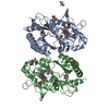
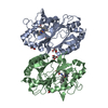
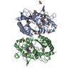
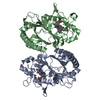
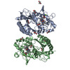
| Title | Dictyostelium discoideum dye decolorizing peroxidase DyPA in complex with cyanide. | ||||||
 Components Components | DyPA | ||||||
 Keywords Keywords | HYDROLASE / Dye decolorizing-type peroxidase / Dictyostelium discoideum / B-type DyP | ||||||
| Function / homology |  Function and homology information Function and homology informationperoxidase activity / protein dimerization activity / heme binding / metal ion binding / cytosol / cytoplasm Similarity search - Function | ||||||
| Biological species |  | ||||||
| Method |  X-RAY DIFFRACTION / SYNCHROTRON / MOLECULAR REPLACEMENT / Resolution: 1.85 Å X-RAY DIFFRACTION / SYNCHROTRON / MOLECULAR REPLACEMENT / Resolution: 1.85 Å | ||||||
 Authors Authors | Rai, A. / Fedorov, R. / Manstein, D.J. | ||||||
| Funding support |  Germany, 1items Germany, 1items
| ||||||
 Citation Citation | Journal: Int J Mol Sci / Year: 2021 Title: Structural and Biochemical Characterization of a Dye-Decolorizing Peroxidase from Dictyostelium discoideum . Authors: Rai, A. / Klare, J.P. / Reinke, P.Y.A. / Englmaier, F. / Fohrer, J. / Fedorov, R. / Taft, M.H. / Chizhov, I. / Curth, U. / Plettenburg, O. / Manstein, D.J. | ||||||
| History |
|
- Structure visualization
| Structure viewer | Molecule: MolmilJmol/JSmol |
|---|
- Downloads & links
-Download
| PDBx/mmCIF format | 7o9l.cif.gz | 328.7 KB | Display | PDBx/mmCIF format |
|---|---|---|---|---|
| PDB format | pdb7o9l.ent.gz | 221.4 KB | Display | PDB format |
| PDBx/mmJSON format | 7o9l.json.gz | Tree view | PDBx/mmJSON format | |
| Others |  Other downloads Other downloads |
-Validation report
| Arichive directory | https://data.pdbj.org/pub/pdb/validation_reports/o9/7o9lftp://data.pdbj.org/pub/pdb/validation_reports/o9/7o9l | HTTPS FTP |
|---|
-Related structure data
| Related structure data |  7o9jSC  7odzC S: Starting model for refinement C: citing same article ( |
|---|---|
| Similar structure data |

-Links
 PDBj
PDBj


- Assembly
| Deposited unit | 
| ||||||||||||
|---|---|---|---|---|---|---|---|---|---|---|---|---|---|
| 1 |
| ||||||||||||
| Unit cell |
|
-Components
| #1: Protein | Mass: 35426.309 Da / Num. of mol.: 2 Source method: isolated from a genetically manipulated source Source: (gene. exp.)  #2: Chemical |   Mass: 616.487 Da / Num. of mol.: 2 / Source method: obtained synthetically / Formula: C34H32FeN4O4 / Feature type: SUBJECT OF INVESTIGATION Mass: 616.487 Da / Num. of mol.: 2 / Source method: obtained synthetically / Formula: C34H32FeN4O4 / Feature type: SUBJECT OF INVESTIGATION#3: Chemical | ChemComp-EDO /   Mass: 62.068 Da / Num. of mol.: 7 / Source method: obtained synthetically / Formula: C2H6O2 Mass: 62.068 Da / Num. of mol.: 7 / Source method: obtained synthetically / Formula: C2H6O2#4: Chemical |   Mass: 26.017 Da / Num. of mol.: 2 / Source method: obtained synthetically / Formula: CN / Feature type: SUBJECT OF INVESTIGATION Mass: 26.017 Da / Num. of mol.: 2 / Source method: obtained synthetically / Formula: CN / Feature type: SUBJECT OF INVESTIGATION#5: Water | ChemComp-HOH / |  Mass: 18.015 Da / Num. of mol.: 393 / Source method: isolated from a natural source / Formula: H2O Mass: 18.015 Da / Num. of mol.: 393 / Source method: isolated from a natural source / Formula: H2OHas ligand of interest | Y | |
|---|
-Experimental details
-Experiment
| Experiment | Method: X-RAY DIFFRACTION / Number of used crystals: 1 |
|---|
- Sample preparation
| Crystal | Density Matthews: 3.35 Å3/Da / Density % sol: 63.3 % |
|---|---|
| Crystal grow | Temperature: 293 K / Method: vapor diffusion / pH: 7 / Details: 2.4 M Sodium malonate pH 7.0 |
-Data collection
| Diffraction | Mean temperature: 100 K / Serial crystal experiment: N |
|---|---|
| Diffraction source | Source: SYNCHROTRON / Site: ESRF  / Beamline: ID29 / Wavelength: 0.91985 Å / Beamline: ID29 / Wavelength: 0.91985 Å |
| Detector | Type: DECTRIS PILATUS 6M / Detector: PIXEL / Date: Dec 1, 2012 |
| Radiation | Monochromator: M / Protocol: SINGLE WAVELENGTH / Monochromatic (M) / Laue (L): M / Scattering type: x-ray |
| Radiation wavelength | Wavelength: 0.91985 Å / Relative weight: 1 |
| Reflection | Resolution: 1.85→47.74 Å / Num. obs: 82374 / % possible obs: 100 % / Redundancy: 26.4 % / Biso Wilson estimate: 25.87 Å2 / Rmerge(I) obs: 0.0394 / Net I/σ(I): 15.36 |
| Reflection shell | Resolution: 1.85→1.94 Å / Redundancy: 26.22 % / Rmerge(I) obs: 0.36 / Num. unique obs: 10792 / % possible all: 100 |
- Processing
| Software |
| ||||||||||||||||||||||||||||||||||||||||||||||||||||||||||||||||||||||||||||||||||||||||||||||||||||||||||||||||||||||||||||||||||||||||||||||||||||||||||||||||||||||||||||||||||||||||||||||||||||||||||||||||||
|---|---|---|---|---|---|---|---|---|---|---|---|---|---|---|---|---|---|---|---|---|---|---|---|---|---|---|---|---|---|---|---|---|---|---|---|---|---|---|---|---|---|---|---|---|---|---|---|---|---|---|---|---|---|---|---|---|---|---|---|---|---|---|---|---|---|---|---|---|---|---|---|---|---|---|---|---|---|---|---|---|---|---|---|---|---|---|---|---|---|---|---|---|---|---|---|---|---|---|---|---|---|---|---|---|---|---|---|---|---|---|---|---|---|---|---|---|---|---|---|---|---|---|---|---|---|---|---|---|---|---|---|---|---|---|---|---|---|---|---|---|---|---|---|---|---|---|---|---|---|---|---|---|---|---|---|---|---|---|---|---|---|---|---|---|---|---|---|---|---|---|---|---|---|---|---|---|---|---|---|---|---|---|---|---|---|---|---|---|---|---|---|---|---|---|---|---|---|---|---|---|---|---|---|---|---|---|---|---|---|---|---|
| Refinement | Method to determine structure: MOLECULAR REPLACEMENT Starting model: 7O9J Resolution: 1.85→45.22 Å / SU ML: 0.1737 / Cross valid method: FREE R-VALUE / σ(F): 1.34 / Phase error: 17.8398 Stereochemistry target values: GeoStd + Monomer Library + CDL v1.2
| ||||||||||||||||||||||||||||||||||||||||||||||||||||||||||||||||||||||||||||||||||||||||||||||||||||||||||||||||||||||||||||||||||||||||||||||||||||||||||||||||||||||||||||||||||||||||||||||||||||||||||||||||||
| Solvent computation | Shrinkage radii: 0.9 Å / VDW probe radii: 1.11 Å / Solvent model: FLAT BULK SOLVENT MODEL | ||||||||||||||||||||||||||||||||||||||||||||||||||||||||||||||||||||||||||||||||||||||||||||||||||||||||||||||||||||||||||||||||||||||||||||||||||||||||||||||||||||||||||||||||||||||||||||||||||||||||||||||||||
| Displacement parameters | Biso mean: 33.08 Å2 | ||||||||||||||||||||||||||||||||||||||||||||||||||||||||||||||||||||||||||||||||||||||||||||||||||||||||||||||||||||||||||||||||||||||||||||||||||||||||||||||||||||||||||||||||||||||||||||||||||||||||||||||||||
| Refinement step | Cycle: LAST / Resolution: 1.85→45.22 Å
| ||||||||||||||||||||||||||||||||||||||||||||||||||||||||||||||||||||||||||||||||||||||||||||||||||||||||||||||||||||||||||||||||||||||||||||||||||||||||||||||||||||||||||||||||||||||||||||||||||||||||||||||||||
| Refine LS restraints |
| ||||||||||||||||||||||||||||||||||||||||||||||||||||||||||||||||||||||||||||||||||||||||||||||||||||||||||||||||||||||||||||||||||||||||||||||||||||||||||||||||||||||||||||||||||||||||||||||||||||||||||||||||||
| LS refinement shell |
| ||||||||||||||||||||||||||||||||||||||||||||||||||||||||||||||||||||||||||||||||||||||||||||||||||||||||||||||||||||||||||||||||||||||||||||||||||||||||||||||||||||||||||||||||||||||||||||||||||||||||||||||||||
| Refinement TLS params. | Method: refined / Origin x: 9.07372623191 Å / Origin y: 26.3872388582 Å / Origin z: 13.8894603174 Å
| ||||||||||||||||||||||||||||||||||||||||||||||||||||||||||||||||||||||||||||||||||||||||||||||||||||||||||||||||||||||||||||||||||||||||||||||||||||||||||||||||||||||||||||||||||||||||||||||||||||||||||||||||||
| Refinement TLS group | Selection details: all |
