 Movie
Movie Controller

[English] 日本語
 Yorodumi
- PDB-7o8n: NmHR light state structure at 7.5 ms (5 - 10 ms) after photoexcit... -
+ Open data

- Basic information
| Entry | Database: PDB / ID: 7o8n | ||||||
|---|---|---|---|---|---|---|---|
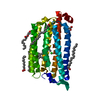
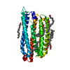
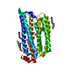
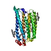
| Title | NmHR light state structure at 7.5 ms (5 - 10 ms) after photoexcitation determined by serial millisecond crystallography | ||||||
 Components Components | Chloride pumping rhodopsin | ||||||
 Keywords Keywords | MEMBRANE PROTEIN / chloride pump / rhodopsin | ||||||
| Function / homology |  Function and homology information Function and homology information | ||||||
| Biological species |  Nonlabens marinus S1-08 (bacteria) Nonlabens marinus S1-08 (bacteria) | ||||||
| Method |  X-RAY DIFFRACTION / SYNCHROTRON / MOLECULAR REPLACEMENT / Resolution: 2.1 Å X-RAY DIFFRACTION / SYNCHROTRON / MOLECULAR REPLACEMENT / Resolution: 2.1 Å | ||||||
 Authors Authors | Mous, S. / Gotthard, G. / Ehrenberg, D. / Sen, S. / James, D. / Johnson, P. / Weinert, T. / Nass, K. / Furrer, A. / Kekilli, D. ...Mous, S. / Gotthard, G. / Ehrenberg, D. / Sen, S. / James, D. / Johnson, P. / Weinert, T. / Nass, K. / Furrer, A. / Kekilli, D. / Ma, P. / Bruenle, S. / Casadei, C. / Martiel, I. / Dworkowski, F. / Gashi, D. / Skopintsev, P. / Wranik, M. / Knopp, G. / Panepucci, E. / Panneels, V. / Cirelli, C. / Ozerov, D. / Schertler, G. / Wang, M. / Milne, C. / Standfuss, J. / Schapiro, I. / Heberle, J. / Nogly, P. | ||||||
| Funding support |  Switzerland, 1items Switzerland, 1items
| ||||||
 Citation Citation | Journal: Science / Year: 2022 Title: Dynamics and mechanism of a light-driven chloride pump. Authors: Mous, S. / Gotthard, G. / Ehrenberg, D. / Sen, S. / Weinert, T. / Johnson, P.J.M. / James, D. / Nass, K. / Furrer, A. / Kekilli, D. / Ma, P. / Brunle, S. / Casadei, C.M. / Martiel, I. / ...Authors: Mous, S. / Gotthard, G. / Ehrenberg, D. / Sen, S. / Weinert, T. / Johnson, P.J.M. / James, D. / Nass, K. / Furrer, A. / Kekilli, D. / Ma, P. / Brunle, S. / Casadei, C.M. / Martiel, I. / Dworkowski, F. / Gashi, D. / Skopintsev, P. / Wranik, M. / Knopp, G. / Panepucci, E. / Panneels, V. / Cirelli, C. / Ozerov, D. / Schertler, G.F.X. / Wang, M. / Milne, C. / Standfuss, J. / Schapiro, I. / Heberle, J. / Nogly, P. | ||||||
| History |
|
- Structure visualization
| Structure viewer | Molecule: MolmilJmol/JSmol |
|---|
- Downloads & links
-Download
| PDBx/mmCIF format | 7o8n.cif.gz | 74.8 KB | Display | PDBx/mmCIF format |
|---|---|---|---|---|
| PDB format | pdb7o8n.ent.gz | 52.4 KB | Display | PDB format |
| PDBx/mmJSON format | 7o8n.json.gz | Tree view | PDBx/mmJSON format | |
| Others |  Other downloads Other downloads |
-Validation report
| Arichive directory | https://data.pdbj.org/pub/pdb/validation_reports/o8/7o8nftp://data.pdbj.org/pub/pdb/validation_reports/o8/7o8n | HTTPS FTP |
|---|
-Related structure data
| Related structure data |  7o8fC  7o8gC  7o8hC  7o8iC  7o8jC  7o8kC  7o8lC  7o8mC  7o8oC  7o8pC  7o8qC  7o8rC  7o8sC  7o8tC  7o8uC  7o8vC  7o8yC  7o8zC  5b2nS S: Starting model for refinement C: citing same article ( |
|---|---|
| Similar structure data |


-Links
 PDBj
PDBj


- Assembly
| Deposited unit | 
| ||||||||
|---|---|---|---|---|---|---|---|---|---|
| 1 |
| ||||||||
| Unit cell |
|
-Components
| #1: Protein | Mass: 32913.434 Da / Num. of mol.: 1 Source method: isolated from a genetically manipulated source Source: (gene. exp.) Nonlabens marinus S1-08 (bacteria) / Gene: ClR, NMS_1267 / Plasmid: pET21b / Production host: | ||||||||
|---|---|---|---|---|---|---|---|---|---|
| #2: Chemical | ChemComp-RET /   Mass: 284.436 Da / Num. of mol.: 1 / Source method: obtained synthetically / Formula: C20H28O Mass: 284.436 Da / Num. of mol.: 1 / Source method: obtained synthetically / Formula: C20H28O | ||||||||
| #3: Chemical |   Mass: 356.540 Da / Num. of mol.: 3 / Source method: obtained synthetically / Formula: C21H40O4 Mass: 356.540 Da / Num. of mol.: 3 / Source method: obtained synthetically / Formula: C21H40O4#4: Chemical | ChemComp-OLA /   Mass: 282.461 Da / Num. of mol.: 6 / Source method: obtained synthetically / Formula: C18H34O2 Mass: 282.461 Da / Num. of mol.: 6 / Source method: obtained synthetically / Formula: C18H34O2#5: Water | ChemComp-HOH / |  Mass: 18.015 Da / Num. of mol.: 50 / Source method: isolated from a natural source / Formula: H2O Mass: 18.015 Da / Num. of mol.: 50 / Source method: isolated from a natural source / Formula: H2OHas ligand of interest | N | Has protein modification | Y | |
-Experimental details
-Experiment
| Experiment | Method: X-RAY DIFFRACTION / Number of used crystals: 1 |
|---|
- Sample preparation
| Crystal | Density Matthews: 2.37 Å3/Da / Density % sol: 48.15 % |
|---|---|
| Crystal grow | Temperature: 293 K / Method: lipidic cubic phase / pH: 4.5 Details: 50 mM sodium acetate (pH 4.5), 150 mM MgCl2, 100 mM NaCl, 34% PEG400 |
-Data collection
| Diffraction | Mean temperature: 298 K / Serial crystal experiment: Y |
|---|---|
| Diffraction source | Source: SYNCHROTRON / Site: SLS / Beamline: X06SA / Wavelength: 1 Å |
| Detector | Type: DECTRIS EIGER X 16M / Detector: PIXEL / Date: May 19, 2019 |
| Radiation | Protocol: SINGLE WAVELENGTH / Monochromatic (M) / Laue (L): M / Scattering type: x-ray |
| Radiation wavelength | Wavelength: 1 Å / Relative weight: 1 |
| Reflection | Resolution: 2→39 Å / Num. obs: 21052 / % possible obs: 100 % / Redundancy: 362 % / Biso Wilson estimate: 36.43 Å2 / CC1/2: 0.99 / Net I/σ(I): 8.5 |
| Reflection shell | Resolution: 2→2.04 Å / Num. unique obs: 2105 / CC1/2: 0.41 |
| Serial crystallography sample delivery | Method: injection |
- Processing
| Software |
| |||||||||||||||||||||||||||||||||||||||||||||||||
|---|---|---|---|---|---|---|---|---|---|---|---|---|---|---|---|---|---|---|---|---|---|---|---|---|---|---|---|---|---|---|---|---|---|---|---|---|---|---|---|---|---|---|---|---|---|---|---|---|---|---|
| Refinement | Method to determine structure: MOLECULAR REPLACEMENT Starting model: 5B2N Resolution: 2.1→38.96 Å / SU ML: 0.42 / Cross valid method: THROUGHOUT / σ(F): -0 / Phase error: 37.47 / Stereochemistry target values: ML
| |||||||||||||||||||||||||||||||||||||||||||||||||
| Solvent computation | Shrinkage radii: 0.9 Å / VDW probe radii: 1.11 Å / Solvent model: FLAT BULK SOLVENT MODEL | |||||||||||||||||||||||||||||||||||||||||||||||||
| Displacement parameters | Biso max: 119.39 Å2 / Biso mean: 44.6217 Å2 / Biso min: 23.9 Å2 | |||||||||||||||||||||||||||||||||||||||||||||||||
| Refinement step | Cycle: final / Resolution: 2.1→38.96 Å
| |||||||||||||||||||||||||||||||||||||||||||||||||
| LS refinement shell | Refine-ID: X-RAY DIFFRACTION / Rfactor Rfree error: 0 / Total num. of bins used: 6
|
