[English] 日本語
 Yorodumi
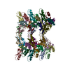
- PDB-7o76: Reversible supramolecular assembly of the anti-microbial peptide ... -
+ Open data
- Basic information
| Entry | Database: PDB / ID: 7o76 | ||||||
|---|---|---|---|---|---|---|---|
| Title | Reversible supramolecular assembly of the anti-microbial peptide plectasin | ||||||
 Components Components | Fungal defensin plectasin | ||||||
 Keywords Keywords | ANTIMICROBIAL PROTEIN / fungal defensin | ||||||
| Function / homology |  Function and homology information Function and homology informationpotassium channel regulator activity / toxin activity / defense response to bacterium / host cell plasma membrane / extracellular region / membrane Similarity search - Function | ||||||
| Biological species |  Pseudoplectania nigrella (fungus) Pseudoplectania nigrella (fungus) | ||||||
| Method |  X-RAY DIFFRACTION / X-RAY DIFFRACTION /  SYNCHROTRON / SYNCHROTRON /  MOLECULAR REPLACEMENT / Resolution: 1.131 Å MOLECULAR REPLACEMENT / Resolution: 1.131 Å | ||||||
 Authors Authors | Pohl, C. / Noergaard, N. / Harris, P. | ||||||
| Funding support | European Union, 1items
| ||||||
 Citation Citation |  Journal: Nat Commun / Year: 2022 Journal: Nat Commun / Year: 2022Title: pH- and concentration-dependent supramolecular assembly of a fungal defensin plectasin variant into helical non-amyloid fibrils. Authors: Christin Pohl / Gregory Effantin / Eaazhisai Kandiah / Sebastian Meier / Guanghong Zeng / Werner Streicher / Dorotea Raventos Segura / Per H Mygind / Dorthe Sandvang / Line Anker Nielsen / ...Authors: Christin Pohl / Gregory Effantin / Eaazhisai Kandiah / Sebastian Meier / Guanghong Zeng / Werner Streicher / Dorotea Raventos Segura / Per H Mygind / Dorthe Sandvang / Line Anker Nielsen / Günther H J Peters / Guy Schoehn / Christoph Mueller-Dieckmann / Allan Noergaard / Pernille Harris /     Abstract: Self-assembly and fibril formation play important roles in protein behaviour. Amyloid fibril formation is well-studied due to its role in neurodegenerative diseases and characterized by refolding of ...Self-assembly and fibril formation play important roles in protein behaviour. Amyloid fibril formation is well-studied due to its role in neurodegenerative diseases and characterized by refolding of the protein into predominantly β-sheet form. However, much less is known about the assembly of proteins into other types of supramolecular structures. Using cryo-electron microscopy at a resolution of 1.97 Å, we show that a triple-mutant of the anti-microbial peptide plectasin, PPI42, assembles into helical non-amyloid fibrils. The in vitro anti-microbial activity was determined and shown to be enhanced compared to the wildtype. Plectasin contains a cysteine-stabilised α-helix-β-sheet structure, which remains intact upon fibril formation. Two protofilaments form a right-handed protein fibril. The fibril formation is reversible and follows sigmoidal kinetics with a pH- and concentration dependent equilibrium between soluble monomer and protein fibril. This high-resolution structure reveals that α/β proteins can natively assemble into fibrils. | ||||||
| History |
|
- Structure visualization
| Structure viewer | Molecule:  Molmil Molmil Jmol/JSmol Jmol/JSmol |
|---|
- Downloads & links
- Download
| PDBx/mmCIF format |  7o76.cif.gz 7o76.cif.gz | 44 KB | Display |  PDBx/mmCIF format PDBx/mmCIF format |
|---|---|---|---|---|
| PDB format |  pdb7o76.ent.gz pdb7o76.ent.gz | Display |  PDB format PDB format | |
| PDBx/mmJSON format |  7o76.json.gz 7o76.json.gz | Tree view |  PDBx/mmJSON format PDBx/mmJSON format | |
| Others |  Other downloads Other downloads |
-Validation report
| Summary document |  7o76_validation.pdf.gz 7o76_validation.pdf.gz | 438.4 KB | Display |  wwPDB validaton report wwPDB validaton report |
|---|---|---|---|---|
| Full document |  7o76_full_validation.pdf.gz 7o76_full_validation.pdf.gz | 438.8 KB | Display | |
| Data in XML |  7o76_validation.xml.gz 7o76_validation.xml.gz | 4.4 KB | Display | |
| Data in CIF |  7o76_validation.cif.gz 7o76_validation.cif.gz | 5.3 KB | Display | |
| Arichive directory |  https://data.pdbj.org/pub/pdb/validation_reports/o7/7o76 https://data.pdbj.org/pub/pdb/validation_reports/o7/7o76 ftp://data.pdbj.org/pub/pdb/validation_reports/o7/7o76 ftp://data.pdbj.org/pub/pdb/validation_reports/o7/7o76 | HTTPS FTP |
-Related structure data
| Related structure data |  7oaeC  7oagC  3e7uS S: Starting model for refinement C: citing same article ( |
|---|---|
| Similar structure data | Similarity search - Function & homology  F&H Search F&H Search |
- Links
Links
- Assembly
| Deposited unit | 
| ||||||||
|---|---|---|---|---|---|---|---|---|---|
| 1 |
| ||||||||
| Unit cell |
|
- Components
| #1: Protein/peptide | Mass: 4415.024 Da / Num. of mol.: 1 Source method: isolated from a genetically manipulated source Source: (gene. exp.)  Pseudoplectania nigrella (fungus) / Gene: DEF / Production host: Pseudoplectania nigrella (fungus) / Gene: DEF / Production host:  |
|---|---|
| #2: Chemical | ChemComp-PEG / |
| #3: Water | ChemComp-HOH / |
| Has ligand of interest | N |
| Has protein modification | Y |
-Experimental details
-Experiment
| Experiment | Method:  X-RAY DIFFRACTION / Number of used crystals: 1 X-RAY DIFFRACTION / Number of used crystals: 1 |
|---|
- Sample preparation
| Crystal | Density Matthews: 1.7 Å3/Da / Density % sol: 27.62 % |
|---|---|
| Crystal grow | Temperature: 300 K / Method: vapor diffusion, hanging drop Details: 15 mg/mL plectasin in 10 mM His pH 4.5 added to a reservoir of 0.1 M NH4Ac, 0.1 M Tris pH 8.5 and 40% isopropanol |
-Data collection
| Diffraction | Mean temperature: 100 K / Serial crystal experiment: N |
|---|---|
| Diffraction source | Source:  SYNCHROTRON / Site: SYNCHROTRON / Site:  MAX IV MAX IV  / Beamline: BioMAX / Wavelength: 0.97247 Å / Beamline: BioMAX / Wavelength: 0.97247 Å |
| Detector | Type: DECTRIS EIGER X 16M / Detector: PIXEL / Date: May 18, 2020 / Details: Kirkpatrick-Baez (KB) mirror pair (VFM, HFM) |
| Radiation | Monochromator: Si (111), double crystal monochromator, horizontally deflecting, LN2 side-cooling Protocol: SINGLE WAVELENGTH / Monochromatic (M) / Laue (L): M / Scattering type: x-ray |
| Radiation wavelength | Wavelength: 0.97247 Å / Relative weight: 1 |
| Reflection | Resolution: 1.13→31.53 Å / Num. obs: 10216 / % possible obs: 89.9 % / Redundancy: 6 % / CC1/2: 0.999 / Net I/σ(I): 22.49 |
| Reflection shell | Resolution: 1.13→1.2 Å / Num. unique obs: 1020 / CC1/2: 0.9 |
- Processing
| Software |
| ||||||||||||||||||||||||||||||||||||||||||||||||||||||||||||||||||||||||||||||||||||||||||||||||||||||||||||||||||||||||||||||||||||||||||||||||||||||
|---|---|---|---|---|---|---|---|---|---|---|---|---|---|---|---|---|---|---|---|---|---|---|---|---|---|---|---|---|---|---|---|---|---|---|---|---|---|---|---|---|---|---|---|---|---|---|---|---|---|---|---|---|---|---|---|---|---|---|---|---|---|---|---|---|---|---|---|---|---|---|---|---|---|---|---|---|---|---|---|---|---|---|---|---|---|---|---|---|---|---|---|---|---|---|---|---|---|---|---|---|---|---|---|---|---|---|---|---|---|---|---|---|---|---|---|---|---|---|---|---|---|---|---|---|---|---|---|---|---|---|---|---|---|---|---|---|---|---|---|---|---|---|---|---|---|---|---|---|---|---|---|
| Refinement | Method to determine structure:  MOLECULAR REPLACEMENT MOLECULAR REPLACEMENTStarting model: 3E7U Resolution: 1.131→21.219 Å / Cor.coef. Fo:Fc: 0.98 / Cor.coef. Fo:Fc free: 0.976 / WRfactor Rfree: 0.215 / WRfactor Rwork: 0.164 / SU B: 2.377 / SU ML: 0.045 / Average fsc free: 0.9365 / Average fsc work: 0.9497 / Cross valid method: FREE R-VALUE / ESU R: 0.04 / ESU R Free: 0.04 Details: Hydrogens have been used if present in the input file
| ||||||||||||||||||||||||||||||||||||||||||||||||||||||||||||||||||||||||||||||||||||||||||||||||||||||||||||||||||||||||||||||||||||||||||||||||||||||
| Solvent computation | Ion probe radii: 0.8 Å / Shrinkage radii: 0.8 Å / VDW probe radii: 1.2 Å / Solvent model: MASK BULK SOLVENT | ||||||||||||||||||||||||||||||||||||||||||||||||||||||||||||||||||||||||||||||||||||||||||||||||||||||||||||||||||||||||||||||||||||||||||||||||||||||
| Displacement parameters | Biso mean: 27.922 Å2
| ||||||||||||||||||||||||||||||||||||||||||||||||||||||||||||||||||||||||||||||||||||||||||||||||||||||||||||||||||||||||||||||||||||||||||||||||||||||
| Refinement step | Cycle: LAST / Resolution: 1.131→21.219 Å
| ||||||||||||||||||||||||||||||||||||||||||||||||||||||||||||||||||||||||||||||||||||||||||||||||||||||||||||||||||||||||||||||||||||||||||||||||||||||
| Refine LS restraints |
| ||||||||||||||||||||||||||||||||||||||||||||||||||||||||||||||||||||||||||||||||||||||||||||||||||||||||||||||||||||||||||||||||||||||||||||||||||||||
| LS refinement shell |
|
 Movie
Movie Controller


 PDBj
PDBj


