 Movie
Movie Controller

[English] 日本語
 Yorodumi
- PDB-7o5q: Crystal Structure of a Class D Carbapenemase Complexed with Hydro... -
+ Open data

- Basic information
| Entry | Database: PDB / ID: 7o5q | ||||||
|---|---|---|---|---|---|---|---|
| Title | Crystal Structure of a Class D Carbapenemase Complexed with Hydrolyzed Oxacillin | ||||||
 Components Components | Beta-lactamase | ||||||
 Keywords Keywords | HYDROLASE / OXA / iodide / oxacillin | ||||||
| Function / homology |  Function and homology information Function and homology informationpenicillin binding / antibiotic catabolic process / cell wall organization / beta-lactamase activity / beta-lactamase / response to antibiotic / plasma membrane Similarity search - Function | ||||||
| Biological species |  Klebsiella pneumoniae (bacteria) Klebsiella pneumoniae (bacteria) | ||||||
| Method |  X-RAY DIFFRACTION / SYNCHROTRON / MOLECULAR REPLACEMENT / Resolution: 1.85 Å X-RAY DIFFRACTION / SYNCHROTRON / MOLECULAR REPLACEMENT / Resolution: 1.85 Å | ||||||
 Authors Authors | Zhou, Q. / He, Y. / Jin, Y. | ||||||
| Funding support |  China, 1items China, 1items
| ||||||
 Citation Citation | Journal: To Be Published Title: Crystal Structure of a Class D Carbapenemase Complexed with Hydrolyzed Oxacillin Authors: Zhou, Q. / He, Y. / Jin, Y. | ||||||
| History |
|
- Structure visualization
| Structure viewer | Molecule: MolmilJmol/JSmol |
|---|
- Downloads & links
-Download
| PDBx/mmCIF format | 7o5q.cif.gz | 418.6 KB | Display | PDBx/mmCIF format |
|---|---|---|---|---|
| PDB format | pdb7o5q.ent.gz | Display | PDB format | |
| PDBx/mmJSON format | 7o5q.json.gz | Tree view | PDBx/mmJSON format | |
| Others |  Other downloads Other downloads |
-Validation report
| Arichive directory | https://data.pdbj.org/pub/pdb/validation_reports/o5/7o5qftp://data.pdbj.org/pub/pdb/validation_reports/o5/7o5q | HTTPS FTP |
|---|
-Related structure data
| Related structure data | 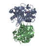 4s2pS S: Starting model for refinement |
|---|---|
| Similar structure data |
-Links
 PDBj
PDBj
- Assembly
| Deposited unit | 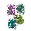
| ||||||||
|---|---|---|---|---|---|---|---|---|---|
| 1 | 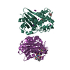
| ||||||||
| 2 | 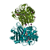
| ||||||||
| Unit cell |
|
-Components
-Protein , 1 types, 4 molecules AAABBBCCCDDD
| #1: Protein | Mass: 30254.133 Da / Num. of mol.: 4 Source method: isolated from a genetically manipulated source Source: (gene. exp.) Klebsiella pneumoniae (bacteria)Gene: bla OXA-48, bla_2, blaOXA-48, G5637_27540, KPE71T_00045, SAMEA3649466_05396 Production host: |
|---|
-Non-polymers , 6 types, 727 molecules 

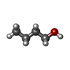
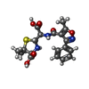
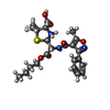

| #2: Chemical |  Mass: 92.094 Da / Num. of mol.: 3 / Source method: obtained synthetically / Formula: C3H8O3 Mass: 92.094 Da / Num. of mol.: 3 / Source method: obtained synthetically / Formula: C3H8O3#3: Chemical | ChemComp-IOD /  Mass: 126.904 Da / Num. of mol.: 15 / Source method: obtained synthetically / Formula: I / Feature type: SUBJECT OF INVESTIGATION Mass: 126.904 Da / Num. of mol.: 15 / Source method: obtained synthetically / Formula: I / Feature type: SUBJECT OF INVESTIGATION#4: Chemical |  Mass: 74.122 Da / Num. of mol.: 2 / Source method: obtained synthetically / Formula: C4H10O Mass: 74.122 Da / Num. of mol.: 2 / Source method: obtained synthetically / Formula: C4H10O#5: Chemical |  Mass: 419.452 Da / Num. of mol.: 2 / Source method: obtained synthetically / Formula: C19H21N3O6S / Feature type: SUBJECT OF INVESTIGATION / Comment: antibiotic*YM Mass: 419.452 Da / Num. of mol.: 2 / Source method: obtained synthetically / Formula: C19H21N3O6S / Feature type: SUBJECT OF INVESTIGATION / Comment: antibiotic*YM#6: Chemical |  Mass: 476.566 Da / Num. of mol.: 2 / Source method: obtained synthetically / Formula: C23H30N3O6S / Feature type: SUBJECT OF INVESTIGATION Mass: 476.566 Da / Num. of mol.: 2 / Source method: obtained synthetically / Formula: C23H30N3O6S / Feature type: SUBJECT OF INVESTIGATION#7: Water | ChemComp-HOH / | Mass: 18.015 Da / Num. of mol.: 703 / Source method: isolated from a natural source / Formula: H2O |
|---|
-Details
| Has ligand of interest | Y |
|---|
-Experimental details
-Experiment
| Experiment | Method: X-RAY DIFFRACTION / Number of used crystals: 1 |
|---|
- Sample preparation
| Crystal | Density Matthews: 2.71 Å3/Da / Density % sol: 54.69 % |
|---|---|
| Crystal grow | Temperature: 291 K / Method: vapor diffusion, hanging drop Details: 0.1M HEPES pH 8.0, 10% PEG 8000, 10% 1-BUTANOL mixed with the 10 mg/mL protein stock at 1:1 ratio. PH range: 7.5-8.0 |
-Data collection
| Diffraction | Mean temperature: 100 K / Serial crystal experiment: N |
|---|---|
| Diffraction source | Source: SYNCHROTRON / Site: SSRF / Beamline: BL17U1 / Wavelength: 0.97915 Å |
| Detector | Type: ADSC QUANTUM 315r / Detector: CCD / Date: Jun 26, 2020 |
| Radiation | Protocol: SINGLE WAVELENGTH / Monochromatic (M) / Laue (L): M / Scattering type: x-ray |
| Radiation wavelength | Wavelength: 0.97915 Å / Relative weight: 1 |
| Reflection | Resolution: 1.85→19.81 Å / Num. obs: 109814 / % possible obs: 99.9 % / Redundancy: 6.9 % / CC1/2: 0.997 / Net I/σ(I): 10 |
| Reflection shell | Resolution: 1.85→1.88 Å / Mean I/σ(I) obs: 1.2 / Num. unique obs: 5493 / CC1/2: 0.725 |
- Processing
| Software |
| |||||||||||||||||||||||||||||||||||||||||||||||||||||||||||||||||||||||||||||||||||||||||||||||||||||||||||||||||||||||||||||||||||||||||||||||||||||||||||
|---|---|---|---|---|---|---|---|---|---|---|---|---|---|---|---|---|---|---|---|---|---|---|---|---|---|---|---|---|---|---|---|---|---|---|---|---|---|---|---|---|---|---|---|---|---|---|---|---|---|---|---|---|---|---|---|---|---|---|---|---|---|---|---|---|---|---|---|---|---|---|---|---|---|---|---|---|---|---|---|---|---|---|---|---|---|---|---|---|---|---|---|---|---|---|---|---|---|---|---|---|---|---|---|---|---|---|---|---|---|---|---|---|---|---|---|---|---|---|---|---|---|---|---|---|---|---|---|---|---|---|---|---|---|---|---|---|---|---|---|---|---|---|---|---|---|---|---|---|---|---|---|---|---|---|---|---|
| Refinement | Method to determine structure: MOLECULAR REPLACEMENT Starting model: 4S2P Resolution: 1.85→19.807 Å / Cor.coef. Fo:Fc: 0.968 / Cor.coef. Fo:Fc free: 0.95 / SU B: 3.851 / SU ML: 0.105 / Cross valid method: FREE R-VALUE / ESU R: 0.117 / ESU R Free: 0.116 Details: Hydrogens have been added in their riding positions
| |||||||||||||||||||||||||||||||||||||||||||||||||||||||||||||||||||||||||||||||||||||||||||||||||||||||||||||||||||||||||||||||||||||||||||||||||||||||||||
| Solvent computation | Ion probe radii: 0.8 Å / Shrinkage radii: 0.8 Å / VDW probe radii: 1.2 Å / Solvent model: MASK BULK SOLVENT | |||||||||||||||||||||||||||||||||||||||||||||||||||||||||||||||||||||||||||||||||||||||||||||||||||||||||||||||||||||||||||||||||||||||||||||||||||||||||||
| Displacement parameters | Biso mean: 27.894 Å2
| |||||||||||||||||||||||||||||||||||||||||||||||||||||||||||||||||||||||||||||||||||||||||||||||||||||||||||||||||||||||||||||||||||||||||||||||||||||||||||
| Refinement step | Cycle: LAST / Resolution: 1.85→19.807 Å
| |||||||||||||||||||||||||||||||||||||||||||||||||||||||||||||||||||||||||||||||||||||||||||||||||||||||||||||||||||||||||||||||||||||||||||||||||||||||||||
| Refine LS restraints |
| |||||||||||||||||||||||||||||||||||||||||||||||||||||||||||||||||||||||||||||||||||||||||||||||||||||||||||||||||||||||||||||||||||||||||||||||||||||||||||
| LS refinement shell |
|
