 Movie
Movie Controller

[English] 日本語
 Yorodumi
- PDB-7nzy: Crystal structure of human Casein Kinase I delta in complex with ... -
+ Open data

- Basic information
| Entry | Database: PDB / ID: 7nzy | ||||||
|---|---|---|---|---|---|---|---|
| Title | Crystal structure of human Casein Kinase I delta in complex with CGS-15943 | ||||||
 Components Components | Casein kinase I isoform delta | ||||||
 Keywords Keywords | TRANSFERASE / Kinase / Inhibitor / Complex / ATP-competitive | ||||||
| Function / homology |  Function and homology information Function and homology informationpositive regulation of non-canonical Wnt signaling pathway / protein localization to Golgi apparatus / COPII vesicle coating / microtubule nucleation / midbrain dopaminergic neuron differentiation / protein localization to cilium / tau-protein kinase / non-motile cilium assembly / protein localization to centrosome / COPII-mediated vesicle transport ...positive regulation of non-canonical Wnt signaling pathway / protein localization to Golgi apparatus / COPII vesicle coating / microtubule nucleation / midbrain dopaminergic neuron differentiation / protein localization to cilium / tau-protein kinase / non-motile cilium assembly / protein localization to centrosome / COPII-mediated vesicle transport / tau-protein kinase activity / Golgi organization / Major pathway of rRNA processing in the nucleolus and cytosol / spindle assembly / endoplasmic reticulum-Golgi intermediate compartment membrane / Loss of Nlp from mitotic centrosomes / Loss of proteins required for interphase microtubule organization from the centrosome / Recruitment of mitotic centrosome proteins and complexes / Recruitment of NuMA to mitotic centrosomes / Anchoring of the basal body to the plasma membrane / AURKA Activation by TPX2 / spindle microtubule / circadian regulation of gene expression / regulation of circadian rhythm / spindle / endocytosis / Wnt signaling pathway / : / Regulation of PLK1 Activity at G2/M Transition / positive regulation of canonical Wnt signaling pathway / positive regulation of proteasomal ubiquitin-dependent protein catabolic process / actin cytoskeleton / protein phosphorylation / protein kinase activity / non-specific serine/threonine protein kinase / cilium / ciliary basal body / cadherin binding / protein serine kinase activity / protein serine/threonine kinase activity / centrosome / perinuclear region of cytoplasm / Golgi apparatus / signal transduction / nucleoplasm / ATP binding / nucleus / plasma membrane / cytosol / cytoplasm Similarity search - Function | ||||||
| Biological species |  Homo sapiens (human) Homo sapiens (human) | ||||||
| Method |  X-RAY DIFFRACTION / SYNCHROTRON / MOLECULAR REPLACEMENT / Resolution: 1.85 Å X-RAY DIFFRACTION / SYNCHROTRON / MOLECULAR REPLACEMENT / Resolution: 1.85 Å | ||||||
 Authors Authors | Pichlo, C. / Baumann, U. | ||||||
 Citation Citation | Journal: J.Med.Chem. / Year: 2022 Title: Phenotypic Discovery of Triazolo[1,5- c ]quinazolines as a First-In-Class Bone Morphogenetic Protein Amplifier Chemotype. Authors: Wesseler, F. / Lohmann, S. / Riege, D. / Halver, J. / Roth, A. / Pichlo, C. / Weber, S. / Takamiya, M. / Muller, E. / Ketzel, J. / Flegel, J. / Gihring, A. / Rastegar, S. / Bertrand, J. / ...Authors: Wesseler, F. / Lohmann, S. / Riege, D. / Halver, J. / Roth, A. / Pichlo, C. / Weber, S. / Takamiya, M. / Muller, E. / Ketzel, J. / Flegel, J. / Gihring, A. / Rastegar, S. / Bertrand, J. / Baumann, U. / Knippschild, U. / Peifer, C. / Sievers, S. / Waldmann, H. / Schade, D. | ||||||
| History |
|
- Structure visualization
| Structure viewer | Molecule: MolmilJmol/JSmol |
|---|
- Downloads & links
-Download
| PDBx/mmCIF format | 7nzy.cif.gz | 513.9 KB | Display | PDBx/mmCIF format |
|---|---|---|---|---|
| PDB format | pdb7nzy.ent.gz | 404.2 KB | Display | PDB format |
| PDBx/mmJSON format | 7nzy.json.gz | Tree view | PDBx/mmJSON format | |
| Others |  Other downloads Other downloads |
-Validation report
| Arichive directory | https://data.pdbj.org/pub/pdb/validation_reports/nz/7nzyftp://data.pdbj.org/pub/pdb/validation_reports/nz/7nzy | HTTPS FTP |
|---|
-Related structure data
| Related structure data | 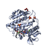 4twcS S: Starting model for refinement |
|---|---|
| Similar structure data |
-Links
 PDBj
PDBj


- Assembly
| Deposited unit | 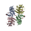
| ||||||||||||
|---|---|---|---|---|---|---|---|---|---|---|---|---|---|
| 1 | 
| ||||||||||||
| 2 | 
| ||||||||||||
| 3 | 
| ||||||||||||
| 4 | 
| ||||||||||||
| Unit cell |
|
-Components
| #1: Protein | Mass: 36457.867 Da / Num. of mol.: 4 Source method: isolated from a genetically manipulated source Source: (gene. exp.) Homo sapiens (human) / Gene: CSNK1D, HCKID / Plasmid: pET28a / Production host:  References: UniProt: P48730, non-specific serine/threonine protein kinase, tau-protein kinase #2: Chemical | ChemComp-UX2 /   Mass: 285.689 Da / Num. of mol.: 4 / Source method: obtained synthetically / Formula: C13H8ClN5O / Feature type: SUBJECT OF INVESTIGATION / Comment: antagonist*YM Mass: 285.689 Da / Num. of mol.: 4 / Source method: obtained synthetically / Formula: C13H8ClN5O / Feature type: SUBJECT OF INVESTIGATION / Comment: antagonist*YM#3: Chemical | ChemComp-MLI /   Mass: 102.046 Da / Num. of mol.: 4 / Source method: obtained synthetically / Formula: C3H2O4 Mass: 102.046 Da / Num. of mol.: 4 / Source method: obtained synthetically / Formula: C3H2O4#4: Chemical | ChemComp-PO4 /   Mass: 94.971 Da / Num. of mol.: 4 / Source method: obtained synthetically / Formula: PO4 Mass: 94.971 Da / Num. of mol.: 4 / Source method: obtained synthetically / Formula: PO4#5: Water | ChemComp-HOH / |  Mass: 18.015 Da / Num. of mol.: 663 / Source method: isolated from a natural source / Formula: H2O Mass: 18.015 Da / Num. of mol.: 663 / Source method: isolated from a natural source / Formula: H2OHas ligand of interest | Y | Has protein modification | Y | |
|---|
-Experimental details
-Experiment
| Experiment | Method: X-RAY DIFFRACTION / Number of used crystals: 1 |
|---|
- Sample preparation
| Crystal | Density Matthews: 2.37 Å3/Da / Density % sol: 48.06 % |
|---|---|
| Crystal grow | Temperature: 294 K / Method: vapor diffusion, sitting drop / Details: 0.2 M sodium malonate pH = 5.0, 20% (w/v) PEG 3350 |
-Data collection
| Diffraction | Mean temperature: 100 K / Serial crystal experiment: N |
|---|---|
| Diffraction source | Source: SYNCHROTRON / Site: PETRA III, EMBL c/o DESY  / Beamline: P13 (MX1) / Wavelength: 0.9794 Å / Beamline: P13 (MX1) / Wavelength: 0.9794 Å |
| Detector | Type: DECTRIS PILATUS 6M / Detector: PIXEL / Date: Jun 15, 2018 |
| Radiation | Protocol: SINGLE WAVELENGTH / Monochromatic (M) / Laue (L): M / Scattering type: x-ray |
| Radiation wavelength | Wavelength: 0.9794 Å / Relative weight: 1 |
| Reflection | Resolution: 1.85→47.04 Å / Num. obs: 110137 / % possible obs: 96.27 % / Redundancy: 3.6 % / Biso Wilson estimate: 28.9 Å2 / CC1/2: 0.997 / Net I/σ(I): 9.96 |
| Reflection shell | Resolution: 1.85→1.916 Å / Num. unique obs: 10873 / CC1/2: 0.797 |
- Processing
| Software |
| ||||||||||||||||||||||||||||||||||||||||||||||||||||||||||||||||||||||||||||||||||||||||||||||||||
|---|---|---|---|---|---|---|---|---|---|---|---|---|---|---|---|---|---|---|---|---|---|---|---|---|---|---|---|---|---|---|---|---|---|---|---|---|---|---|---|---|---|---|---|---|---|---|---|---|---|---|---|---|---|---|---|---|---|---|---|---|---|---|---|---|---|---|---|---|---|---|---|---|---|---|---|---|---|---|---|---|---|---|---|---|---|---|---|---|---|---|---|---|---|---|---|---|---|---|---|
| Refinement | Method to determine structure: MOLECULAR REPLACEMENT Starting model: 4TWC Resolution: 1.85→47.04 Å / SU ML: 0.2443 / Cross valid method: FREE R-VALUE / σ(F): 2.03 / Phase error: 30.9771 Stereochemistry target values: GeoStd + Monomer Library + CDL v1.2
| ||||||||||||||||||||||||||||||||||||||||||||||||||||||||||||||||||||||||||||||||||||||||||||||||||
| Solvent computation | Shrinkage radii: 0.9 Å / VDW probe radii: 1.11 Å / Solvent model: FLAT BULK SOLVENT MODEL | ||||||||||||||||||||||||||||||||||||||||||||||||||||||||||||||||||||||||||||||||||||||||||||||||||
| Displacement parameters | Biso mean: 44.93 Å2 | ||||||||||||||||||||||||||||||||||||||||||||||||||||||||||||||||||||||||||||||||||||||||||||||||||
| Refinement step | Cycle: LAST / Resolution: 1.85→47.04 Å
| ||||||||||||||||||||||||||||||||||||||||||||||||||||||||||||||||||||||||||||||||||||||||||||||||||
| Refine LS restraints |
| ||||||||||||||||||||||||||||||||||||||||||||||||||||||||||||||||||||||||||||||||||||||||||||||||||
| LS refinement shell |
|
