 Movie
Movie Controller


+ Open data

- Basic information
| Entry | Database: PDB / ID: 7ntn | ||||||
|---|---|---|---|---|---|---|---|
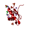
| Title | The structure of RRM domain of human TRMT2A at 2 A resolution | ||||||
 Components Components | tRNA (uracil-5-)-methyltransferase homolog A | ||||||
 Keywords Keywords | TRANSFERASE / tRNA binding / methyltransferase / TRMT2a / polyQ aggregation | ||||||
| Function / homology |  Function and homology information Function and homology informationtRNA (uracil(54)-C5)-methyltransferase activity, S-adenosyl methionine-dependent / tRNA (uracil54-C5)-methyltransferase / C-methyltransferase activity / tRNA processing / Transferases; Transferring one-carbon groups; Methyltransferases / mRNA processing / methylation / RNA binding / cytosol Similarity search - Function | ||||||
| Biological species |  Homo sapiens (human) Homo sapiens (human) | ||||||
| Method |  X-RAY DIFFRACTION / SYNCHROTRON / MOLECULAR REPLACEMENT / Resolution: 2.016 Å X-RAY DIFFRACTION / SYNCHROTRON / MOLECULAR REPLACEMENT / Resolution: 2.016 Å | ||||||
 Authors Authors | Davydova, E. / Janowski, R. / Witzenberger, M. / Niessing, D. | ||||||
| Funding support |  Germany, 1items Germany, 1items
| ||||||
 Citation Citation | Journal: Comput Struct Biotechnol J / Year: 2022 Title: Small-molecule modulators of TRMT2A decrease PolyQ aggregation and PolyQ-induced cell death. Authors: Margreiter, M.A. / Witzenberger, M. / Wasser, Y. / Davydova, E. / Janowski, R. / Metz, J. / Habib, P. / Sahnoun, S.E.M. / Sobisch, C. / Poma, B. / Palomino-Hernandez, O. / Wagner, M. / ...Authors: Margreiter, M.A. / Witzenberger, M. / Wasser, Y. / Davydova, E. / Janowski, R. / Metz, J. / Habib, P. / Sahnoun, S.E.M. / Sobisch, C. / Poma, B. / Palomino-Hernandez, O. / Wagner, M. / Carell, T. / Jon Shah, N. / Schulz, J.B. / Niessing, D. / Voigt, A. / Rossetti, G. | ||||||
| History |
|
- Structure visualization
| Structure viewer | Molecule: MolmilJmol/JSmol |
|---|
- Downloads & links
-Download
| PDBx/mmCIF format | 7ntn.cif.gz | 50.9 KB | Display | PDBx/mmCIF format |
|---|---|---|---|---|
| PDB format | pdb7ntn.ent.gz | 37 KB | Display | PDB format |
| PDBx/mmJSON format | 7ntn.json.gz | Tree view | PDBx/mmJSON format | |
| Others |  Other downloads Other downloads |
-Validation report
| Summary document | 7ntn_validation.pdf.gz | 433.6 KB | Display | wwPDB validaton report |
|---|---|---|---|---|
| Full document | 7ntn_full_validation.pdf.gz | 433.5 KB | Display | |
| Data in XML | 7ntn_validation.xml.gz | 5.7 KB | Display | |
| Data in CIF | 7ntn_validation.cif.gz | 7 KB | Display | |
| Arichive directory | https://data.pdbj.org/pub/pdb/validation_reports/nt/7ntnftp://data.pdbj.org/pub/pdb/validation_reports/nt/7ntn | HTTPS FTP |
-Related structure data


-Links
 PDBj
PDBj- Assembly
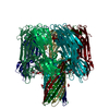
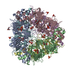
| Deposited unit | 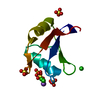
| |||||||||||||||
|---|---|---|---|---|---|---|---|---|---|---|---|---|---|---|---|---|
| 1 | x 24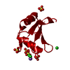
| |||||||||||||||
| Unit cell |
| |||||||||||||||
| Components on special symmetry positions |
|
-Components
| #1: Protein | Mass: 9213.796 Da / Num. of mol.: 1 Source method: isolated from a genetically manipulated source Source: (gene. exp.) Homo sapiens (human) / Gene: TRMT2A, HTF9C / Production host:  References: UniProt: Q8IZ69, Transferases; Transferring one-carbon groups; Methyltransferases | ||||||||
|---|---|---|---|---|---|---|---|---|---|
| #2: Chemical | ChemComp-SO4 /   Mass: 96.063 Da / Num. of mol.: 4 / Source method: obtained synthetically / Formula: SO4 Mass: 96.063 Da / Num. of mol.: 4 / Source method: obtained synthetically / Formula: SO4#3: Chemical |   Mass: 35.453 Da / Num. of mol.: 3 / Source method: obtained synthetically / Formula: Cl Mass: 35.453 Da / Num. of mol.: 3 / Source method: obtained synthetically / Formula: Cl#4: Chemical | ChemComp-NA / |   Mass: 22.990 Da / Num. of mol.: 1 / Source method: obtained synthetically / Formula: Na Mass: 22.990 Da / Num. of mol.: 1 / Source method: obtained synthetically / Formula: Na#5: Water | ChemComp-HOH / |  Mass: 18.015 Da / Num. of mol.: 31 / Source method: isolated from a natural source / Formula: H2O Mass: 18.015 Da / Num. of mol.: 31 / Source method: isolated from a natural source / Formula: H2OHas ligand of interest | N | |
-Experimental details
-Experiment
| Experiment | Method: X-RAY DIFFRACTION / Number of used crystals: 1 |
|---|
- Sample preparation
| Crystal | Density Matthews: 2.57 Å3/Da / Density % sol: 52.1 % |
|---|---|
| Crystal grow | Temperature: 292 K / Method: vapor diffusion, hanging drop / pH: 4.5 Details: 0.22M lithium sulfate, 0.1M sodium acetate (pH 4.5) and 26 % PEG 6000 |
-Data collection
| Diffraction | Mean temperature: 100 K / Serial crystal experiment: N |
|---|---|
| Diffraction source | Source: SYNCHROTRON / Site: PETRA III, DESY / Beamline: P11 / Wavelength: 1.0093 Å |
| Detector | Type: DECTRIS PILATUS 6M-F / Detector: PIXEL / Date: Jul 16, 2016 |
| Radiation | Protocol: SINGLE WAVELENGTH / Monochromatic (M) / Laue (L): M / Scattering type: x-ray |
| Radiation wavelength | Wavelength: 1.0093 Å / Relative weight: 1 |
| Reflection | Resolution: 2.016→50 Å / Num. obs: 6872 / % possible obs: 100 % / Redundancy: 29.45 % / CC1/2: 1 / Net I/σ(I): 36.42 |
| Reflection shell | Resolution: 2.016→2.07 Å / Mean I/σ(I) obs: 3.85 / Num. unique obs: 1079 / CC1/2: 0.92 |
- Processing
| Software |
| ||||||||||||||||||||||||||||||||||||||||
|---|---|---|---|---|---|---|---|---|---|---|---|---|---|---|---|---|---|---|---|---|---|---|---|---|---|---|---|---|---|---|---|---|---|---|---|---|---|---|---|---|---|
| Refinement | Method to determine structure: MOLECULAR REPLACEMENT Starting model: sequence Resolution: 2.016→46.475 Å / SU ML: 0.23 / Cross valid method: THROUGHOUT / σ(F): 1.35 / Phase error: 28.73 / Stereochemistry target values: ML
| ||||||||||||||||||||||||||||||||||||||||
| Solvent computation | Shrinkage radii: 0.9 Å / VDW probe radii: 1.11 Å / Solvent model: FLAT BULK SOLVENT MODEL | ||||||||||||||||||||||||||||||||||||||||
| Displacement parameters | Biso max: 160.09 Å2 / Biso mean: 52.1131 Å2 / Biso min: 24.3 Å2 | ||||||||||||||||||||||||||||||||||||||||
| Refinement step | Cycle: final / Resolution: 2.016→46.475 Å
| ||||||||||||||||||||||||||||||||||||||||
| Refine LS restraints |
| ||||||||||||||||||||||||||||||||||||||||
| LS refinement shell | Refine-ID: X-RAY DIFFRACTION / Rfactor Rfree error: 0 / % reflection obs: 100 %
| ||||||||||||||||||||||||||||||||||||||||
| Refinement TLS params. | Method: refined / Origin x: 55.576 Å / Origin y: 34.0871 Å / Origin z: 15.2121 Å
| ||||||||||||||||||||||||||||||||||||||||
| Refinement TLS group |
|
