 Movie
Movie Controller

[English] 日本語
 Yorodumi
- PDB-7nru: Structure of a natural chimera of meningococcal factor H binding ... -
+ Open data

- Basic information
| Entry | Database: PDB / ID: 7nru | ||||||
|---|---|---|---|---|---|---|---|
| Title | Structure of a natural chimera of meningococcal factor H binding protein belonging to NL096 strain | ||||||
 Components Components | Factor H binding protein variant 1-2,3.x | ||||||
 Keywords Keywords | PROTEIN BINDING / meningococcal antigen / natural chimera | ||||||
| Function / homology | Factor H binding protein, C-terminal / : / : / Factor H binding protein, C-terminal / Factor H binding protein, N-terminal / Outer membrane protein/outer membrane enzyme PagP, beta-barrel / cell outer membrane / Factor H binding protein variant A62_001 Function and homology information Function and homology information | ||||||
| Biological species |  Neisseria meningitidis (bacteria) Neisseria meningitidis (bacteria) | ||||||
| Method |  X-RAY DIFFRACTION / SYNCHROTRON / MOLECULAR REPLACEMENT / Resolution: 1.21998357323 Å X-RAY DIFFRACTION / SYNCHROTRON / MOLECULAR REPLACEMENT / Resolution: 1.21998357323 Å | ||||||
 Authors Authors | Veggi, D. / Malito, E. / Bottomley, M.J. | ||||||
 Citation Citation | Journal: Comput Struct Biotechnol J / Year: 2022 Title: Structural characterization of a cross-protective natural chimera of factor H binding protein from meningococcal serogroup B strain NL096. Authors: Veggi, D. / Malito, E. / Lo Surdo, P. / Pansegrau, W. / Rippa, V. / Wahome, N. / Savino, S. / Masignani, V. / Pizza, M. / Bottomley, M.J. #1: Journal: Patent / Year: 2020Title: Neisseria meningitidis compositions and methods thereof Authors: Jansen, K.U. / Anderson, A.S. / Absalon, J. / Aste-Amezaga, J.M. / Beeslaar, J.F. / Cooper, D. / Farley, J.E. / Fletcher, L.D. / Harris, S.L. / Jones, T.R. / Kanevsky, I. / Khandke, L. / ...Authors: Jansen, K.U. / Anderson, A.S. / Absalon, J. / Aste-Amezaga, J.M. / Beeslaar, J.F. / Cooper, D. / Farley, J.E. / Fletcher, L.D. / Harris, S.L. / Jones, T.R. / Kanevsky, I. / Khandke, L. / Liberator, P. / Perez, J.L. / Phelan, L.M. / Zlotnick, G.W. | ||||||
| History |
|
- Structure visualization
| Structure viewer | Molecule: MolmilJmol/JSmol |
|---|
- Downloads & links
-Download
| PDBx/mmCIF format | 7nru.cif.gz | 158.5 KB | Display | PDBx/mmCIF format |
|---|---|---|---|---|
| PDB format | pdb7nru.ent.gz | 121.4 KB | Display | PDB format |
| PDBx/mmJSON format | 7nru.json.gz | Tree view | PDBx/mmJSON format | |
| Others |  Other downloads Other downloads |
-Validation report
| Summary document | 7nru_validation.pdf.gz | 431.2 KB | Display | wwPDB validaton report |
|---|---|---|---|---|
| Full document | 7nru_full_validation.pdf.gz | 432.9 KB | Display | |
| Data in XML | 7nru_validation.xml.gz | 13.7 KB | Display | |
| Data in CIF | 7nru_validation.cif.gz | 20.5 KB | Display | |
| Arichive directory | https://data.pdbj.org/pub/pdb/validation_reports/nr/7nruftp://data.pdbj.org/pub/pdb/validation_reports/nr/7nru | HTTPS FTP |
-Related structure data
| Related structure data |  3kvdS S: Starting model for refinement |
|---|---|
| Similar structure data |
-Links
 PDBj
PDBj

- Assembly
| Deposited unit | 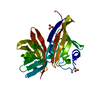
| ||||||||||||
|---|---|---|---|---|---|---|---|---|---|---|---|---|---|
| 1 |
| ||||||||||||
| Unit cell |
|
-Components
| #1: Protein | Mass: 27900.188 Da / Num. of mol.: 1 Source method: isolated from a genetically manipulated source Source: (gene. exp.) Neisseria meningitidis (bacteria) / Gene: fhbp / Production host: | ||||
|---|---|---|---|---|---|
| #2: Chemical |   Mass: 96.063 Da / Num. of mol.: 2 / Source method: isolated from a natural source / Formula: SO4 Mass: 96.063 Da / Num. of mol.: 2 / Source method: isolated from a natural source / Formula: SO4#3: Water | ChemComp-HOH / |  Mass: 18.015 Da / Num. of mol.: 264 / Source method: isolated from a natural source / Formula: H2O Mass: 18.015 Da / Num. of mol.: 264 / Source method: isolated from a natural source / Formula: H2OHas ligand of interest | N | |
-Experimental details
-Experiment
| Experiment | Method: X-RAY DIFFRACTION / Number of used crystals: 1 |
|---|
- Sample preparation
| Crystal | Density Matthews: 2.1 Å3/Da / Density % sol: 41.35 % |
|---|---|
| Crystal grow | Temperature: 294.15 K / Method: vapor diffusion, sitting drop / pH: 5 / Details: 0.1M tri-sodium citrate, 3.2M ammonium sulfate |
-Data collection
| Diffraction | Mean temperature: 100 K / Serial crystal experiment: N |
|---|---|
| Diffraction source | Source: SYNCHROTRON / Site: SLS  / Beamline: X06DA / Wavelength: 0.99987 Å / Beamline: X06DA / Wavelength: 0.99987 Å |
| Detector | Type: DECTRIS PILATUS 2M / Detector: PIXEL / Date: Jan 30, 2012 |
| Radiation | Protocol: SINGLE WAVELENGTH / Monochromatic (M) / Laue (L): M / Scattering type: x-ray |
| Radiation wavelength | Wavelength: 0.99987 Å / Relative weight: 1 |
| Reflection | Resolution: 1.219→23.37 Å / Num. obs: 65693 / % possible obs: 95.2 % / Redundancy: 3 % / Biso Wilson estimate: 10.1827524357 Å2 / Rmerge(I) obs: 0.063 / Net I/σ(I): 10.5 |
| Reflection shell | Resolution: 1.22→1.264 Å / Rmerge(I) obs: 0.2 / Mean I/σ(I) obs: 4.4 / Num. unique obs: 9280 |
- Processing
| Software |
| |||||||||||||||||||||||||||||||||||||||||||||||||||||||||||||||||||||||||||||||||||||||||||||||||||||||||||||||||||||||||||||||||||||||||||||||||||||||||||||||||||||||||||||||
|---|---|---|---|---|---|---|---|---|---|---|---|---|---|---|---|---|---|---|---|---|---|---|---|---|---|---|---|---|---|---|---|---|---|---|---|---|---|---|---|---|---|---|---|---|---|---|---|---|---|---|---|---|---|---|---|---|---|---|---|---|---|---|---|---|---|---|---|---|---|---|---|---|---|---|---|---|---|---|---|---|---|---|---|---|---|---|---|---|---|---|---|---|---|---|---|---|---|---|---|---|---|---|---|---|---|---|---|---|---|---|---|---|---|---|---|---|---|---|---|---|---|---|---|---|---|---|---|---|---|---|---|---|---|---|---|---|---|---|---|---|---|---|---|---|---|---|---|---|---|---|---|---|---|---|---|---|---|---|---|---|---|---|---|---|---|---|---|---|---|---|---|---|---|---|---|---|
| Refinement | Method to determine structure: MOLECULAR REPLACEMENT Starting model: 3KVD Resolution: 1.21998357323→23.3683816098 Å / SU ML: 0.0836274587195 / Cross valid method: FREE R-VALUE / σ(F): 1.3571381526 / Phase error: 15.1064659749 Stereochemistry target values: GeoStd + Monomer Library + CDL v1.2
| |||||||||||||||||||||||||||||||||||||||||||||||||||||||||||||||||||||||||||||||||||||||||||||||||||||||||||||||||||||||||||||||||||||||||||||||||||||||||||||||||||||||||||||||
| Solvent computation | Shrinkage radii: 0.9 Å / VDW probe radii: 1.11 Å / Solvent model: FLAT BULK SOLVENT MODEL | |||||||||||||||||||||||||||||||||||||||||||||||||||||||||||||||||||||||||||||||||||||||||||||||||||||||||||||||||||||||||||||||||||||||||||||||||||||||||||||||||||||||||||||||
| Displacement parameters | Biso mean: 16.576031317 Å2 | |||||||||||||||||||||||||||||||||||||||||||||||||||||||||||||||||||||||||||||||||||||||||||||||||||||||||||||||||||||||||||||||||||||||||||||||||||||||||||||||||||||||||||||||
| Refinement step | Cycle: LAST / Resolution: 1.21998357323→23.3683816098 Å
| |||||||||||||||||||||||||||||||||||||||||||||||||||||||||||||||||||||||||||||||||||||||||||||||||||||||||||||||||||||||||||||||||||||||||||||||||||||||||||||||||||||||||||||||
| Refine LS restraints |
| |||||||||||||||||||||||||||||||||||||||||||||||||||||||||||||||||||||||||||||||||||||||||||||||||||||||||||||||||||||||||||||||||||||||||||||||||||||||||||||||||||||||||||||||
| LS refinement shell |
|
