 Movie
Movie Controller

[English] 日本語
 Yorodumi
- PDB-7nox: Structure of SGBP BO2743 from Bacteroides ovatus in complex with ... -
+ Open data

- Basic information
| Entry | Database: PDB / ID: 7nox | |||||||||
|---|---|---|---|---|---|---|---|---|---|---|
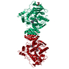
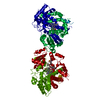
| Title | Structure of SGBP BO2743 from Bacteroides ovatus in complex with mixed-linked gluco-nonasaccharide | |||||||||
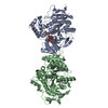
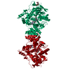
 Components Components | Surface glycan-binding protein BO2743 | |||||||||
 Keywords Keywords | SUGAR BINDING PROTEIN / Bacteroides ovatus / surface glycan binding protein (SGBP) / SusD-homologue / Polysaccharide Utilization Loci (PUL) / gut microbiota / beta-glucans / barley-9 | |||||||||
| Function / homology | SusD-like 2 / Starch-binding associating with outer membrane / Prokaryotic membrane lipoprotein lipid attachment site profile. / Tetratricopeptide-like helical domain superfamily / AZIDE ION / TRIETHYLENE GLYCOL / SusD/RagB family nutrient-binding outer membrane lipoprotein Function and homology information Function and homology information | |||||||||
| Biological species |  Bacteroides ovatus ATCC 8483 (bacteria) Bacteroides ovatus ATCC 8483 (bacteria) | |||||||||
| Method |  X-RAY DIFFRACTION / SYNCHROTRON / MOLECULAR REPLACEMENT / Resolution: 1.43 Å X-RAY DIFFRACTION / SYNCHROTRON / MOLECULAR REPLACEMENT / Resolution: 1.43 Å | |||||||||
 Authors Authors | Correia, V.C. / Trovao, F. / Pinheiro, B.A. / Palma, A.S. / Carvalho, A.L. | |||||||||
| Funding support |  Portugal, 2items Portugal, 2items
| |||||||||
 Citation Citation | Journal: Microbiol Spectr / Year: 2021 Title: Mapping Molecular Recognition of beta 1,3-1,4-Glucans by a Surface Glycan-Binding Protein from the Human Gut Symbiont Bacteroides ovatus. Authors: Correia, V.G. / Trovao, F. / Pinheiro, B.A. / Bras, J.L.A. / Silva, L.M. / Nunes, C. / Coimbra, M.A. / Liu, Y. / Feizi, T. / Fontes, C.M.G.A. / Mulloy, B. / Chai, W. / Carvalho, A.L. / Palma, A.S. | |||||||||
| History |
|
- Structure visualization
| Structure viewer | Molecule: MolmilJmol/JSmol |
|---|
- Downloads & links
-Download
| PDBx/mmCIF format | 7nox.cif.gz | 254.3 KB | Display | PDBx/mmCIF format |
|---|---|---|---|---|
| PDB format | pdb7nox.ent.gz | 199.4 KB | Display | PDB format |
| PDBx/mmJSON format | 7nox.json.gz | Tree view | PDBx/mmJSON format | |
| Others |  Other downloads Other downloads |
-Validation report
| Summary document | 7nox_validation.pdf.gz | 941 KB | Display | wwPDB validaton report |
|---|---|---|---|---|
| Full document | 7nox_full_validation.pdf.gz | 946.2 KB | Display | |
| Data in XML | 7nox_validation.xml.gz | 46.9 KB | Display | |
| Data in CIF | 7nox_validation.cif.gz | 72.7 KB | Display | |
| Arichive directory | https://data.pdbj.org/pub/pdb/validation_reports/no/7noxftp://data.pdbj.org/pub/pdb/validation_reports/no/7nox | HTTPS FTP |
-Related structure data
| Related structure data | 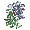 7o8cC 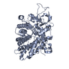 5j5uS S: Starting model for refinement C: citing same article ( |
|---|---|
| Similar structure data |

-Links
 PDBj
PDBj


- Assembly
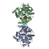
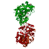
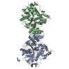
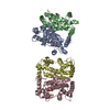
| Deposited unit | 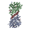
| ||||||||
|---|---|---|---|---|---|---|---|---|---|
| 1 |
| ||||||||
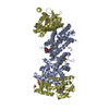
| Unit cell |
|
-Components
-Protein / Sugars , 2 types, 4 molecules AB
| #1: Protein | Mass: 67918.438 Da / Num. of mol.: 2 Source method: isolated from a genetically manipulated source Source: (gene. exp.) Bacteroides ovatus ATCC 8483 (bacteria)Gene: BACOVA_02743 / Production host: #2: Polysaccharide | Type: oligosaccharide / Mass: 1477.282 Da / Num. of mol.: 2 Source method: isolated from a genetically manipulated source |
|---|
-Non-polymers , 7 types, 1023 molecules 


| #3: Chemical |  Mass: 24.305 Da / Num. of mol.: 2 / Source method: obtained synthetically / Formula: Mg Mass: 24.305 Da / Num. of mol.: 2 / Source method: obtained synthetically / Formula: Mg#4: Chemical | ChemComp-GOL /  Mass: 92.094 Da / Num. of mol.: 5 / Source method: obtained synthetically / Formula: C3H8O3 Mass: 92.094 Da / Num. of mol.: 5 / Source method: obtained synthetically / Formula: C3H8O3#5: Chemical | ChemComp-PGE /  Mass: 150.173 Da / Num. of mol.: 8 / Source method: obtained synthetically / Formula: C6H14O4 Mass: 150.173 Da / Num. of mol.: 8 / Source method: obtained synthetically / Formula: C6H14O4#6: Chemical |  Mass: 22.990 Da / Num. of mol.: 3 / Source method: obtained synthetically / Formula: Na Mass: 22.990 Da / Num. of mol.: 3 / Source method: obtained synthetically / Formula: Na#7: Chemical | ChemComp-AZI /  Mass: 42.020 Da / Num. of mol.: 14 / Source method: obtained synthetically / Formula: N3 Mass: 42.020 Da / Num. of mol.: 14 / Source method: obtained synthetically / Formula: N3#8: Chemical | ChemComp-TAM / |  Mass: 163.215 Da / Num. of mol.: 1 / Source method: obtained synthetically / Formula: C7H17NO3 / Comment: pH buffer*YM Mass: 163.215 Da / Num. of mol.: 1 / Source method: obtained synthetically / Formula: C7H17NO3 / Comment: pH buffer*YM#9: Water | ChemComp-HOH / | Mass: 18.015 Da / Num. of mol.: 990 / Source method: isolated from a natural source / Formula: H2O |
|---|
-Details
| Has ligand of interest | Y |
|---|
-Experimental details
-Experiment
| Experiment | Method: X-RAY DIFFRACTION / Number of used crystals: 1 |
|---|
- Sample preparation
| Crystal | Density Matthews: 2.24 Å3/Da / Density % sol: 45 % |
|---|---|
| Crystal grow | Temperature: 293 K / Method: vapor diffusion, sitting drop Details: Protein at 8 mg/mL; 25% PEG 3350, 0.1 M Bis-Tris pH 5.5, 0.2 mM MgCl2.6H2O |
-Data collection
| Diffraction | Mean temperature: 100 K / Serial crystal experiment: N |
|---|---|
| Diffraction source | Source: SYNCHROTRON / Site: Diamond  / Beamline: I02 / Wavelength: 0.9795 Å / Beamline: I02 / Wavelength: 0.9795 Å |
| Detector | Type: DECTRIS PILATUS 6M-F / Detector: PIXEL / Date: Jul 26, 2019 |
| Radiation | Protocol: SINGLE WAVELENGTH / Monochromatic (M) / Laue (L): M / Scattering type: x-ray |
| Radiation wavelength | Wavelength: 0.9795 Å / Relative weight: 1 |
| Reflection | Resolution: 1.43→62.32 Å / Num. obs: 175289 / % possible obs: 96.5 % / Redundancy: 9 % / CC1/2: 1 / Net I/σ(I): 1.6 |
| Reflection shell | Resolution: 1.43→1.55 Å / Num. unique obs: 8764 / CC1/2: 0.74 |
- Processing
| Software |
| ||||||||||||||||||
|---|---|---|---|---|---|---|---|---|---|---|---|---|---|---|---|---|---|---|---|
| Refinement | Method to determine structure: MOLECULAR REPLACEMENT Starting model: 5J5U Resolution: 1.43→44.86 Å / Cross valid method: THROUGHOUT
| ||||||||||||||||||
| Displacement parameters | Biso max: 80.83 Å2 / Biso mean: 23.0947 Å2 / Biso min: 10.51 Å2 | ||||||||||||||||||
| Refinement step | Cycle: LAST / Resolution: 1.43→44.86 Å
| ||||||||||||||||||
| LS refinement shell | Resolution: 1.43→1.45 Å
|
