 Movie
Movie Controller


+ Open data

- Basic information
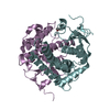
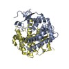
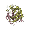
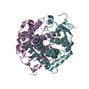
| Entry | Database: PDB / ID: 7nc8 | ||||||
|---|---|---|---|---|---|---|---|
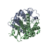
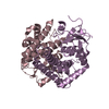
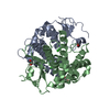
| Title | Glutathione-S-transferase GliG mutant S24A | ||||||
 Components Components | Glutathione S-transferase GliG | ||||||
 Keywords Keywords | BIOSYNTHETIC PROTEIN / Aspergillus fumigatus / mycotoxin / glutathione-S-transferase / carbon-sulphur-bond / epidithiodioxopiperazine | ||||||
| Function / homology |  Function and homology information Function and homology informationglutathione transferase / glutathione transferase activity / molecular function activator activity Similarity search - Function | ||||||
| Biological species |  | ||||||
| Method |  X-RAY DIFFRACTION / SYNCHROTRON / MOLECULAR REPLACEMENT / Resolution: 2.2 Å X-RAY DIFFRACTION / SYNCHROTRON / MOLECULAR REPLACEMENT / Resolution: 2.2 Å | ||||||
 Authors Authors | Groll, M. / Huber, E.M. | ||||||
 Citation Citation | Journal: Angew.Chem.Int.Ed.Engl. / Year: 2021 Title: Structural and Mechanistic Insights into C-S Bond Formation in Gliotoxin. Authors: Scherlach, K. / Kuttenlochner, W. / Scharf, D.H. / Brakhage, A.A. / Hertweck, C. / Groll, M. / Huber, E.M. | ||||||
| History |
|
- Structure visualization
| Structure viewer | Molecule: MolmilJmol/JSmol |
|---|
- Downloads & links
-Download
| PDBx/mmCIF format | 7nc8.cif.gz | 590.6 KB | Display | PDBx/mmCIF format |
|---|---|---|---|---|
| PDB format | pdb7nc8.ent.gz | 491.2 KB | Display | PDB format |
| PDBx/mmJSON format | 7nc8.json.gz | Tree view | PDBx/mmJSON format | |
| Others |  Other downloads Other downloads |
-Validation report
| Arichive directory | https://data.pdbj.org/pub/pdb/validation_reports/nc/7nc8ftp://data.pdbj.org/pub/pdb/validation_reports/nc/7nc8 | HTTPS FTP |
|---|
-Related structure data
| Related structure data | 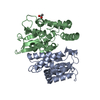 7nc1C 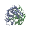 7nc2C  7nc3SC  7nc5C  7nc6C  7nc9C 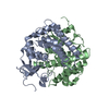 7ncbC 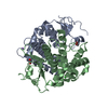 7ncdC 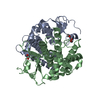 7nceC  7nclC 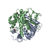 7ncmC 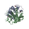 7ncnC  7ncoC  7ncpC  7nctC 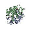 7ncuC S: Starting model for refinement C: citing same article ( |
|---|---|
| Similar structure data |
-Links
 PDBj
PDBj

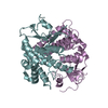
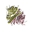
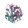
- Assembly
| Deposited unit | 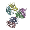
| ||||||||
|---|---|---|---|---|---|---|---|---|---|
| 1 | 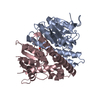
| ||||||||
| 2 | 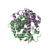
| ||||||||
| 3 | 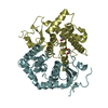
| ||||||||
| Unit cell |
|
-Components
| #1: Protein | Mass: 28947.014 Da / Num. of mol.: 6 / Mutation: S24A Source method: isolated from a genetically manipulated source Source: (gene. exp.) Strain: CEA10 / CBS 144.89 / FGSC A1163 / Gene: AFUB_075740 / Production host:  #2: Chemical | ChemComp-EDO /   Mass: 62.068 Da / Num. of mol.: 6 / Source method: obtained synthetically / Formula: C2H6O2 Mass: 62.068 Da / Num. of mol.: 6 / Source method: obtained synthetically / Formula: C2H6O2#3: Water | ChemComp-HOH / |  Mass: 18.015 Da / Num. of mol.: 438 / Source method: isolated from a natural source / Formula: H2O Mass: 18.015 Da / Num. of mol.: 438 / Source method: isolated from a natural source / Formula: H2OHas ligand of interest | N | |
|---|
-Experimental details
-Experiment
| Experiment | Method: X-RAY DIFFRACTION / Number of used crystals: 1 |
|---|
- Sample preparation
| Crystal | Density Matthews: 2.54 Å3/Da / Density % sol: 51.53 % |
|---|---|
| Crystal grow | Temperature: 293 K / Method: vapor diffusion, sitting drop / pH: 5.5 Details: 0.2 M ammonium acetate, 0.1 M bis-tris pH 5.5, 25 % PEG3350 |
-Data collection
| Diffraction | Mean temperature: 100 K / Serial crystal experiment: N |
|---|---|
| Diffraction source | Source: SYNCHROTRON / Site: SLS  / Beamline: X06SA / Wavelength: 1 Å / Beamline: X06SA / Wavelength: 1 Å |
| Detector | Type: DECTRIS EIGER X 16M / Detector: PIXEL / Date: Nov 4, 2017 |
| Radiation | Protocol: SINGLE WAVELENGTH / Monochromatic (M) / Laue (L): M / Scattering type: x-ray |
| Radiation wavelength | Wavelength: 1 Å / Relative weight: 1 |
| Reflection | Resolution: 2.2→49 Å / Num. obs: 89697 / % possible obs: 99.2 % / Redundancy: 4.2 % / Rmerge(I) obs: 0.053 / Net I/σ(I): 15.3 |
| Reflection shell | Resolution: 2.2→2.3 Å / Rmerge(I) obs: 0.533 / Mean I/σ(I) obs: 2.8 / Num. unique obs: 11149 |
- Processing
| Software |
| |||||||||||||||||||||||||||||||||||||||||||||||||||||||||||||||||||||||||||||||||||||||||||||||||||||||||||||||||||||||||||||||||||||||||||||||||||||||||||||||||||||||||||||||
|---|---|---|---|---|---|---|---|---|---|---|---|---|---|---|---|---|---|---|---|---|---|---|---|---|---|---|---|---|---|---|---|---|---|---|---|---|---|---|---|---|---|---|---|---|---|---|---|---|---|---|---|---|---|---|---|---|---|---|---|---|---|---|---|---|---|---|---|---|---|---|---|---|---|---|---|---|---|---|---|---|---|---|---|---|---|---|---|---|---|---|---|---|---|---|---|---|---|---|---|---|---|---|---|---|---|---|---|---|---|---|---|---|---|---|---|---|---|---|---|---|---|---|---|---|---|---|---|---|---|---|---|---|---|---|---|---|---|---|---|---|---|---|---|---|---|---|---|---|---|---|---|---|---|---|---|---|---|---|---|---|---|---|---|---|---|---|---|---|---|---|---|---|---|---|---|---|
| Refinement | Method to determine structure: MOLECULAR REPLACEMENT Starting model: 7NC3 Resolution: 2.2→30 Å / Cor.coef. Fo:Fc: 0.958 / Cor.coef. Fo:Fc free: 0.935 / SU B: 14.073 / SU ML: 0.155 / Cross valid method: THROUGHOUT / σ(F): 0 / ESU R Free: 0.199 / Stereochemistry target values: MAXIMUM LIKELIHOOD Details: HYDROGENS HAVE BEEN ADDED IN THE RIDING POSITIONS U VALUES : WITH TLS ADDED
| |||||||||||||||||||||||||||||||||||||||||||||||||||||||||||||||||||||||||||||||||||||||||||||||||||||||||||||||||||||||||||||||||||||||||||||||||||||||||||||||||||||||||||||||
| Solvent computation | Ion probe radii: 0.8 Å / Shrinkage radii: 0.8 Å / VDW probe radii: 1.2 Å / Solvent model: MASK | |||||||||||||||||||||||||||||||||||||||||||||||||||||||||||||||||||||||||||||||||||||||||||||||||||||||||||||||||||||||||||||||||||||||||||||||||||||||||||||||||||||||||||||||
| Displacement parameters | Biso max: 144.79 Å2 / Biso mean: 51.679 Å2 / Biso min: 26.78 Å2
| |||||||||||||||||||||||||||||||||||||||||||||||||||||||||||||||||||||||||||||||||||||||||||||||||||||||||||||||||||||||||||||||||||||||||||||||||||||||||||||||||||||||||||||||
| Refinement step | Cycle: final / Resolution: 2.2→30 Å
| |||||||||||||||||||||||||||||||||||||||||||||||||||||||||||||||||||||||||||||||||||||||||||||||||||||||||||||||||||||||||||||||||||||||||||||||||||||||||||||||||||||||||||||||
| Refine LS restraints |
| |||||||||||||||||||||||||||||||||||||||||||||||||||||||||||||||||||||||||||||||||||||||||||||||||||||||||||||||||||||||||||||||||||||||||||||||||||||||||||||||||||||||||||||||
| LS refinement shell | Resolution: 2.2→2.257 Å / Rfactor Rfree error: 0 / Total num. of bins used: 20
| |||||||||||||||||||||||||||||||||||||||||||||||||||||||||||||||||||||||||||||||||||||||||||||||||||||||||||||||||||||||||||||||||||||||||||||||||||||||||||||||||||||||||||||||
| Refinement TLS params. | Method: refined / Refine-ID: X-RAY DIFFRACTION
| |||||||||||||||||||||||||||||||||||||||||||||||||||||||||||||||||||||||||||||||||||||||||||||||||||||||||||||||||||||||||||||||||||||||||||||||||||||||||||||||||||||||||||||||
| Refinement TLS group |
|
