 Movie
Movie Controller

[English] 日本語
 Yorodumi
- PDB-7n5n: PCNA from Thermococcus gammatolerans: crystal III, collection 15,... -
+ Open data

- Basic information
| Entry | Database: PDB / ID: 7n5n | ||||||
|---|---|---|---|---|---|---|---|
| Title | PCNA from Thermococcus gammatolerans: crystal III, collection 15, 2.20 A, 28.7 MGy | ||||||
 Components Components | DNA polymerase sliding clamp | ||||||
 Keywords Keywords | DNA BINDING PROTEIN / PCNA / sliding clamp / radioresistance / radiation damage / ionizing radiation | ||||||
| Function / homology |  Function and homology information Function and homology informationDNA polymerase processivity factor activity / leading strand elongation / regulation of DNA replication / DNA binding Similarity search - Function | ||||||
| Biological species |   Thermococcus gammatolerans (archaea) Thermococcus gammatolerans (archaea) | ||||||
| Method |  X-RAY DIFFRACTION / SYNCHROTRON / MOLECULAR REPLACEMENT / Resolution: 2.2 Å X-RAY DIFFRACTION / SYNCHROTRON / MOLECULAR REPLACEMENT / Resolution: 2.2 Å | ||||||
 Authors Authors | Marin-Tovar, Y. / Rudino-Pinera, E. | ||||||
| Funding support |  Mexico, 1items Mexico, 1items
| ||||||
 Citation Citation | Journal: Proteins / Year: 2022 Title: PCNA from Thermococcus gammatolerans: A protein involved in chromosomal DNA metabolism intrinsically resistant at high levels of ionizing radiation. Authors: Marin-Tovar, Y. / Serrano-Posada, H. / Diaz-Vilchis, A. / Rudino-Pinera, E. | ||||||
| History |
|
- Structure visualization
| Structure viewer | Molecule: MolmilJmol/JSmol |
|---|
- Downloads & links
-Download
| PDBx/mmCIF format | 7n5n.cif.gz | 117 KB | Display | PDBx/mmCIF format |
|---|---|---|---|---|
| PDB format | pdb7n5n.ent.gz | 89.9 KB | Display | PDB format |
| PDBx/mmJSON format | 7n5n.json.gz | Tree view | PDBx/mmJSON format | |
| Others |  Other downloads Other downloads |
-Validation report
| Arichive directory | https://data.pdbj.org/pub/pdb/validation_reports/n5/7n5nftp://data.pdbj.org/pub/pdb/validation_reports/n5/7n5n | HTTPS FTP |
|---|
-Related structure data
| Related structure data |  7n5iC  7n5jC  7n5kC  7n5lC  7n5mC  5a6dS S: Starting model for refinement C: citing same article ( |
|---|---|
| Similar structure data |
-Links
 PDBj
PDBj

- Assembly
| Deposited unit | 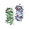
| ||||||||
|---|---|---|---|---|---|---|---|---|---|
| 1 | 
| ||||||||
| 2 | 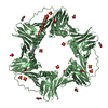
| ||||||||
| Unit cell |
|
-Components
| #1: Protein | Mass: 30191.521 Da / Num. of mol.: 2 Source method: isolated from a genetically manipulated source Source: (gene. exp.) Thermococcus gammatolerans (strain DSM 15229 / JCM 11827 / EJ3) (archaea)Strain: DSM 15229 / JCM 11827 / EJ3 / Gene: pcn, TGAM_1046 / Plasmid: pCold-I / Production host:  #2: Chemical | ChemComp-GOL /   Mass: 92.094 Da / Num. of mol.: 8 / Source method: obtained synthetically / Formula: C3H8O3 Mass: 92.094 Da / Num. of mol.: 8 / Source method: obtained synthetically / Formula: C3H8O3#3: Chemical | ChemComp-SO4 /   Mass: 96.063 Da / Num. of mol.: 8 / Source method: obtained synthetically / Formula: SO4 Mass: 96.063 Da / Num. of mol.: 8 / Source method: obtained synthetically / Formula: SO4#4: Water | ChemComp-HOH / |  Mass: 18.015 Da / Num. of mol.: 125 / Source method: isolated from a natural source / Formula: H2O Mass: 18.015 Da / Num. of mol.: 125 / Source method: isolated from a natural source / Formula: H2OHas ligand of interest | N | |
|---|
-Experimental details
-Experiment
| Experiment | Method: X-RAY DIFFRACTION / Number of used crystals: 1 |
|---|
- Sample preparation
| Crystal | Density Matthews: 2.65 Å3/Da / Density % sol: 54 % |
|---|---|
| Crystal grow | Temperature: 291 K / Method: microbatch / pH: 5.2 Details: 100 mM sodium citrate pH 5.2, 3.0 M ammonium sulfate, 7.5% MPD |
-Data collection
| Diffraction | Mean temperature: 100 K / Serial crystal experiment: N |
|---|---|
| Diffraction source | Source: SYNCHROTRON / Site: SSRL  / Beamline: BL12-2 / Wavelength: 0.9795 Å / Beamline: BL12-2 / Wavelength: 0.9795 Å |
| Detector | Type: DECTRIS PILATUS 6M / Detector: PIXEL / Date: Apr 25, 2018 |
| Radiation | Protocol: SINGLE WAVELENGTH / Monochromatic (M) / Laue (L): M / Scattering type: x-ray |
| Radiation wavelength | Wavelength: 0.9795 Å / Relative weight: 1 |
| Reflection | Resolution: 2.2→34.095 Å / Num. obs: 31432 / % possible obs: 99.62 % / Redundancy: 5.1 % / CC1/2: 0.999 / Rmerge(I) obs: 0.1307 / Net I/σ(I): 19.02 |
| Reflection shell | Resolution: 2.2→2.28 Å / Redundancy: 5.1 % / Rmerge(I) obs: 0.986 / Mean I/σ(I) obs: 2.37 / Num. unique obs: 3150 / CC1/2: 0.748 / % possible all: 99.75 |
- Processing
| Software |
| ||||||||||||||||||||||||||||||||||||||||||||||||||||||||||||||||||||||||
|---|---|---|---|---|---|---|---|---|---|---|---|---|---|---|---|---|---|---|---|---|---|---|---|---|---|---|---|---|---|---|---|---|---|---|---|---|---|---|---|---|---|---|---|---|---|---|---|---|---|---|---|---|---|---|---|---|---|---|---|---|---|---|---|---|---|---|---|---|---|---|---|---|---|
| Refinement | Method to determine structure: MOLECULAR REPLACEMENT Starting model: 5A6D Resolution: 2.2→34.095 Å / SU ML: 0.25 / Cross valid method: THROUGHOUT / σ(F): 1.96 / Phase error: 24.04 / Stereochemistry target values: ML
| ||||||||||||||||||||||||||||||||||||||||||||||||||||||||||||||||||||||||
| Solvent computation | Shrinkage radii: 0.9 Å / VDW probe radii: 1.11 Å / Solvent model: FLAT BULK SOLVENT MODEL | ||||||||||||||||||||||||||||||||||||||||||||||||||||||||||||||||||||||||
| Displacement parameters | Biso max: 131.75 Å2 / Biso mean: 60.8378 Å2 / Biso min: 34.55 Å2 | ||||||||||||||||||||||||||||||||||||||||||||||||||||||||||||||||||||||||
| Refinement step | Cycle: final / Resolution: 2.2→34.095 Å
| ||||||||||||||||||||||||||||||||||||||||||||||||||||||||||||||||||||||||
| LS refinement shell | Refine-ID: X-RAY DIFFRACTION / Rfactor Rfree error: 0
|
