 Movie
Movie Controller


+ Open data

- Basic information
| Entry | Database: PDB / ID: 7n36 | ||||||
|---|---|---|---|---|---|---|---|
| Title | Crystal structure of wild-type human gamma(S)-crystallin | ||||||
 Components Components | Gamma-crystallin S | ||||||
 Keywords Keywords | STRUCTURAL PROTEIN / crystallin / human crystallin / gamma(S) crystallin / eye lens protein | ||||||
| Function / homology |  Function and homology information Function and homology informationstructural constituent of eye lens / lens development in camera-type eye / visual perception / morphogenesis of an epithelium Similarity search - Function | ||||||
| Biological species |  Homo sapiens (human) Homo sapiens (human) | ||||||
| Method |  X-RAY DIFFRACTION / SYNCHROTRON / MOLECULAR REPLACEMENT / Resolution: 2 Å X-RAY DIFFRACTION / SYNCHROTRON / MOLECULAR REPLACEMENT / Resolution: 2 Å | ||||||
 Authors Authors | Norton-Baker, B. / Mehrabi, P. / Martin, R.W. | ||||||
 Citation Citation | Journal: Structure / Year: 2022 Title: Deamidation of the human eye lens protein gamma S-crystallin accelerates oxidative aging. Authors: Norton-Baker, B. / Mehrabi, P. / Kwok, A.O. / Roskamp, K.W. / Rocha, M.A. / Sprague-Piercy, M.A. / von Stetten, D. / Miller, R.J.D. / Martin, R.W. | ||||||
| History |
|
- Structure visualization
| Structure viewer | Molecule: MolmilJmol/JSmol |
|---|
- Downloads & links
-Download
| PDBx/mmCIF format | 7n36.cif.gz | 88.5 KB | Display | PDBx/mmCIF format |
|---|---|---|---|---|
| PDB format | pdb7n36.ent.gz | 64.6 KB | Display | PDB format |
| PDBx/mmJSON format | 7n36.json.gz | Tree view | PDBx/mmJSON format | |
| Others |  Other downloads Other downloads |
-Validation report
| Arichive directory | https://data.pdbj.org/pub/pdb/validation_reports/n3/7n36ftp://data.pdbj.org/pub/pdb/validation_reports/n3/7n36 | HTTPS FTP |
|---|
-Related structure data
| Related structure data | 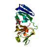 7n37C  7n38C  7n39C 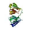 7n3aC  7n3bC 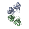 6fd8S S: Starting model for refinement C: citing same article ( |
|---|---|
| Similar structure data |
-Links
 PDBj
PDBj
- Assembly
| Deposited unit | 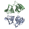
| ||||||||||||
|---|---|---|---|---|---|---|---|---|---|---|---|---|---|
| 1 | 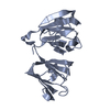
| ||||||||||||
| 2 | 
| ||||||||||||
| Unit cell |
|
-Components
| #1: Protein | Mass: 20959.633 Da / Num. of mol.: 2 Source method: isolated from a genetically manipulated source Source: (gene. exp.) Homo sapiens (human) / Gene: CRYGS, CRYG8 / Production host:  #2: Water | ChemComp-HOH / |  Mass: 18.015 Da / Num. of mol.: 118 / Source method: isolated from a natural source / Formula: H2O Mass: 18.015 Da / Num. of mol.: 118 / Source method: isolated from a natural source / Formula: H2O |
|---|
-Experimental details
-Experiment
| Experiment | Method: X-RAY DIFFRACTION / Number of used crystals: 1 |
|---|
- Sample preparation
| Crystal | Density Matthews: 1.9 Å3/Da / Density % sol: 35.26 % |
|---|---|
| Crystal grow | Temperature: 293 K / Method: vapor diffusion, hanging drop Details: Morpheus condition G5 (0.1 M Carboxylic Acids Mix, 0.1 M Buffer System 2 pH 7.5, 30 % v/v Precipitant Mix 1) |
-Data collection
| Diffraction | Mean temperature: 100 K / Serial crystal experiment: N |
|---|---|
| Diffraction source | Source: SYNCHROTRON / Site: PETRA III, EMBL c/o DESY  / Beamline: P14 (MX2) / Wavelength: 0.9762 Å / Beamline: P14 (MX2) / Wavelength: 0.9762 Å |
| Detector | Type: DECTRIS EIGER X 16M / Detector: PIXEL / Date: Aug 25, 2020 |
| Radiation | Protocol: SINGLE WAVELENGTH / Monochromatic (M) / Laue (L): M / Scattering type: x-ray |
| Radiation wavelength | Wavelength: 0.9762 Å / Relative weight: 1 |
| Reflection | Resolution: 2→46.89 Å / Num. obs: 20807 / % possible obs: 97.42 % / Redundancy: 7 % / Biso Wilson estimate: 39.6 Å2 / CC1/2: 0.999 / Net I/σ(I): 16.29 |
| Reflection shell | Resolution: 2→2.07 Å / Num. unique obs: 2056 / CC1/2: 0.782 |
- Processing
| Software |
| ||||||||||||||||||||||||||||||||||||||||||||||||||||||||
|---|---|---|---|---|---|---|---|---|---|---|---|---|---|---|---|---|---|---|---|---|---|---|---|---|---|---|---|---|---|---|---|---|---|---|---|---|---|---|---|---|---|---|---|---|---|---|---|---|---|---|---|---|---|---|---|---|---|
| Refinement | Method to determine structure: MOLECULAR REPLACEMENT Starting model: 6FD8 Resolution: 2→46.89 Å / SU ML: 0.2381 / Cross valid method: FREE R-VALUE / σ(F): 1.36 / Phase error: 26.9133 Stereochemistry target values: GeoStd + Monomer Library + CDL v1.2
| ||||||||||||||||||||||||||||||||||||||||||||||||||||||||
| Solvent computation | Shrinkage radii: 0.9 Å / VDW probe radii: 1.11 Å / Solvent model: FLAT BULK SOLVENT MODEL | ||||||||||||||||||||||||||||||||||||||||||||||||||||||||
| Displacement parameters | Biso mean: 43.59 Å2 | ||||||||||||||||||||||||||||||||||||||||||||||||||||||||
| Refinement step | Cycle: LAST / Resolution: 2→46.89 Å
| ||||||||||||||||||||||||||||||||||||||||||||||||||||||||
| Refine LS restraints |
| ||||||||||||||||||||||||||||||||||||||||||||||||||||||||
| LS refinement shell |
|
