 Movie
Movie Controller

[English] 日本語
 Yorodumi
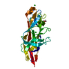
- PDB-7mzs: Crystal structure of the UcaD lectin-binding domain in complex wi... -
+ Open data

- Basic information
| Entry | Database: PDB / ID: 7mzs | ||||||
|---|---|---|---|---|---|---|---|
| Title | Crystal structure of the UcaD lectin-binding domain in complex with galactose | ||||||
 Components Components | Fimbrial adhesin UcaD | ||||||
 Keywords Keywords | SUGAR BINDING PROTEIN / lectin / cell adhesion | ||||||
| Function / homology | Fimbrial-type adhesion domain / Fimbrial protein / : / Fimbrial-type adhesion domain superfamily / cell adhesion involved in single-species biofilm formation / Adhesion domain superfamily / pilus / alpha-D-galactopyranose / Fimbrial adhesin Function and homology information Function and homology information | ||||||
| Biological species |  Proteus mirabilis (bacteria) Proteus mirabilis (bacteria) | ||||||
| Method |  X-RAY DIFFRACTION / SYNCHROTRON / MOLECULAR REPLACEMENT / Resolution: 1.72 Å X-RAY DIFFRACTION / SYNCHROTRON / MOLECULAR REPLACEMENT / Resolution: 1.72 Å | ||||||
 Authors Authors | Ve, T. / Lo, A.W. / Schembri, M.A. / Kobe, B. | ||||||
| Funding support |  Australia, 1items Australia, 1items
| ||||||
 Citation Citation | Journal: Plos Pathog. / Year: 2022 Title: Ucl fimbriae regulation and glycan receptor specificity contribute to gut colonisation by extra-intestinal pathogenic Escherichia coli. Authors: Hancock, S.J. / Lo, A.W. / Ve, T. / Day, C.J. / Tan, L. / Mendez, A.A. / Phan, M.D. / Nhu, N.T.K. / Peters, K.M. / Richards, A.C. / Fleming, B.A. / Chang, C. / Ngu, D.H.Y. / Forde, B.M. / ...Authors: Hancock, S.J. / Lo, A.W. / Ve, T. / Day, C.J. / Tan, L. / Mendez, A.A. / Phan, M.D. / Nhu, N.T.K. / Peters, K.M. / Richards, A.C. / Fleming, B.A. / Chang, C. / Ngu, D.H.Y. / Forde, B.M. / Haselhorst, T. / Goh, K.G.K. / Beatson, S.A. / Jennings, M.P. / Mulvey, M.A. / Kobe, B. / Schembri, M.A. | ||||||
| History |
|
- Structure visualization
| Structure viewer | Molecule: MolmilJmol/JSmol |
|---|
- Downloads & links
-Download
| PDBx/mmCIF format | 7mzs.cif.gz | 106.8 KB | Display | PDBx/mmCIF format |
|---|---|---|---|---|
| PDB format | pdb7mzs.ent.gz | 66 KB | Display | PDB format |
| PDBx/mmJSON format | 7mzs.json.gz | Tree view | PDBx/mmJSON format | |
| Others |  Other downloads Other downloads |
-Validation report
| Summary document | 7mzs_validation.pdf.gz | 717.5 KB | Display | wwPDB validaton report |
|---|---|---|---|---|
| Full document | 7mzs_full_validation.pdf.gz | 717.4 KB | Display | |
| Data in XML | 7mzs_validation.xml.gz | 11.2 KB | Display | |
| Data in CIF | 7mzs_validation.cif.gz | 16.7 KB | Display | |
| Arichive directory | https://data.pdbj.org/pub/pdb/validation_reports/mz/7mzsftp://data.pdbj.org/pub/pdb/validation_reports/mz/7mzs | HTTPS FTP |
-Related structure data


-Links
 PDBj
PDBj- Assembly
| Deposited unit | 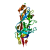
| ||||||||||||
|---|---|---|---|---|---|---|---|---|---|---|---|---|---|
| 1 |
| ||||||||||||
| Unit cell |
| ||||||||||||
| Components on special symmetry positions |
|
-Components
| #1: Protein | Mass: 21985.908 Da / Num. of mol.: 1 Source method: isolated from a genetically manipulated source Source: (gene. exp.) Proteus mirabilis (bacteria) / Gene: NCTC10975_02625 / Production host: | ||||
|---|---|---|---|---|---|
| #2: Sugar | ChemComp-GLA / 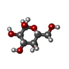  Type: D-saccharide, alpha linking / Mass: 180.156 Da / Num. of mol.: 1 / Source method: obtained synthetically / Formula: C6H12O6 / Feature type: SUBJECT OF INVESTIGATION Type: D-saccharide, alpha linking / Mass: 180.156 Da / Num. of mol.: 1 / Source method: obtained synthetically / Formula: C6H12O6 / Feature type: SUBJECT OF INVESTIGATION | ||||
| #3: Chemical | ChemComp-CL /   Mass: 35.453 Da / Num. of mol.: 6 / Source method: obtained synthetically / Formula: Cl Mass: 35.453 Da / Num. of mol.: 6 / Source method: obtained synthetically / Formula: Cl#4: Water | ChemComp-HOH / |  Mass: 18.015 Da / Num. of mol.: 236 / Source method: isolated from a natural source / Formula: H2O Mass: 18.015 Da / Num. of mol.: 236 / Source method: isolated from a natural source / Formula: H2OHas ligand of interest | Y | |
-Experimental details
-Experiment
| Experiment | Method: X-RAY DIFFRACTION / Number of used crystals: 1 |
|---|
- Sample preparation
| Crystal | Density Matthews: 2.68 Å3/Da / Density % sol: 54.07 % |
|---|---|
| Crystal grow | Temperature: 293 K / Method: vapor diffusion, hanging drop / Details: 0.1 M sodium citrate buffer pH 4.5-5.5, 2-3 M NaCl / PH range: 4.5-5.5 |
-Data collection
| Diffraction | Mean temperature: 100 K / Serial crystal experiment: N |
|---|---|
| Diffraction source | Source: SYNCHROTRON / Site: Australian Synchrotron / Beamline: MX2 / Wavelength: 0.9537 Å |
| Detector | Type: ADSC QUANTUM 315r / Detector: CCD / Date: Jul 30, 2016 |
| Radiation | Protocol: SINGLE WAVELENGTH / Monochromatic (M) / Laue (L): M / Scattering type: x-ray |
| Radiation wavelength | Wavelength: 0.9537 Å / Relative weight: 1 |
| Reflection | Resolution: 1.72→56.12 Å / Num. obs: 23071 / % possible obs: 99.4 % / Redundancy: 3.6 % / Biso Wilson estimate: 13.04 Å2 / CC1/2: 0.99 / Rmerge(I) obs: 0.128 / Rpim(I) all: 0.077 / Rrim(I) all: 0.151 / Net I/σ(I): 8.4 |
| Reflection shell | Resolution: 1.72→1.75 Å / Rmerge(I) obs: 1.114 / Mean I/σ(I) obs: 1.5 / Num. unique obs: 1237 / CC1/2: 0.291 / Rpim(I) all: 0.687 / Rrim(I) all: 1.319 |
- Processing
| Software |
| |||||||||||||||||||||||||||||||||||||||||||||||||||||||||||||||
|---|---|---|---|---|---|---|---|---|---|---|---|---|---|---|---|---|---|---|---|---|---|---|---|---|---|---|---|---|---|---|---|---|---|---|---|---|---|---|---|---|---|---|---|---|---|---|---|---|---|---|---|---|---|---|---|---|---|---|---|---|---|---|---|---|
| Refinement | Method to determine structure: MOLECULAR REPLACEMENT Starting model: UcaD Resolution: 1.72→39.68 Å / SU ML: 0.191 / Cross valid method: FREE R-VALUE / σ(F): 1.35 / Phase error: 22.1686 Stereochemistry target values: GeoStd + Monomer Library + CDL v1.2
| |||||||||||||||||||||||||||||||||||||||||||||||||||||||||||||||
| Solvent computation | Shrinkage radii: 0.9 Å / VDW probe radii: 1.11 Å / Solvent model: FLAT BULK SOLVENT MODEL | |||||||||||||||||||||||||||||||||||||||||||||||||||||||||||||||
| Displacement parameters | Biso mean: 16.55 Å2 | |||||||||||||||||||||||||||||||||||||||||||||||||||||||||||||||
| Refinement step | Cycle: LAST / Resolution: 1.72→39.68 Å
| |||||||||||||||||||||||||||||||||||||||||||||||||||||||||||||||
| Refine LS restraints |
| |||||||||||||||||||||||||||||||||||||||||||||||||||||||||||||||
| LS refinement shell |
|
