 Movie
Movie Controller


+ Open data

- Basic information
| Entry | Database: PDB / ID: 7myy | ||||||
|---|---|---|---|---|---|---|---|
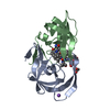
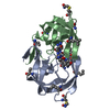
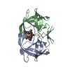
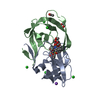
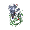
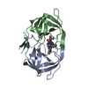
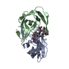
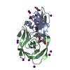
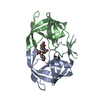
| Title | Crystal Structure of HIV-1 PRS17 with GRL-142 | ||||||
 Components Components | Protease | ||||||
 Keywords Keywords | HYDROLASE/Inhibitor / HIV-1 Protease / Multi drug resistant / Hydrolase inhibitor complex / HYDROLASE / HYDROLASE-Inhibitor complex | ||||||
| Function / homology |  Function and homology information Function and homology informationhost multivesicular body / aspartic-type endopeptidase activity / virion membrane / proteolysis Similarity search - Function | ||||||
| Biological species |   Human immunodeficiency virus 1 Human immunodeficiency virus 1 | ||||||
| Method |  X-RAY DIFFRACTION / SYNCHROTRON / MOLECULAR REPLACEMENT / Resolution: 1.5 Å X-RAY DIFFRACTION / SYNCHROTRON / MOLECULAR REPLACEMENT / Resolution: 1.5 Å | ||||||
 Authors Authors | Agniswamy, J. / Weber, I.T. | ||||||
| Funding support |  United States, 1items United States, 1items
| ||||||
 Citation Citation | Journal: Biochem.Biophys.Res.Commun. / Year: 2021 Title: Novel HIV PR inhibitors with C4-substituted bis-THF and bis-fluoro-benzyl target the two active site mutations of highly drug resistant mutant PR S17 . Authors: Agniswamy, J. / Kneller, D.W. / Ghosh, A.K. / Weber, I.T. | ||||||
| History |
|
- Structure visualization
| Structure viewer | Molecule: MolmilJmol/JSmol |
|---|
- Downloads & links
-Download
| PDBx/mmCIF format | 7myy.cif.gz | 61.4 KB | Display | PDBx/mmCIF format |
|---|---|---|---|---|
| PDB format | pdb7myy.ent.gz | 43.8 KB | Display | PDB format |
| PDBx/mmJSON format | 7myy.json.gz | Tree view | PDBx/mmJSON format | |
| Others |  Other downloads Other downloads |
-Validation report
| Arichive directory | https://data.pdbj.org/pub/pdb/validation_reports/my/7myyftp://data.pdbj.org/pub/pdb/validation_reports/my/7myy | HTTPS FTP |
|---|
-Related structure data
| Related structure data |  7mypC  5t2zS S: Starting model for refinement C: citing same article ( |
|---|---|
| Similar structure data |


-Links
 PDBj
PDBj
- Assembly
| Deposited unit | 
| ||||||||
|---|---|---|---|---|---|---|---|---|---|
| 1 |
| ||||||||
| Unit cell |
|
-Components
| #1: Protein | Mass: 10792.645 Da / Num. of mol.: 2 Source method: isolated from a genetically manipulated source Source: (gene. exp.) Human immunodeficiency virus 1 / Gene: pol / Production host:  #2: Chemical | ChemComp-7OA / ( |   Mass: 706.820 Da / Num. of mol.: 1 / Source method: obtained synthetically / Formula: C33H40F2N4O7S2 / Feature type: SUBJECT OF INVESTIGATION Mass: 706.820 Da / Num. of mol.: 1 / Source method: obtained synthetically / Formula: C33H40F2N4O7S2 / Feature type: SUBJECT OF INVESTIGATION#3: Chemical |   Mass: 92.094 Da / Num. of mol.: 2 / Source method: obtained synthetically / Formula: C3H8O3 Mass: 92.094 Da / Num. of mol.: 2 / Source method: obtained synthetically / Formula: C3H8O3#4: Chemical | ChemComp-CL / |   Mass: 35.453 Da / Num. of mol.: 1 / Source method: obtained synthetically / Formula: Cl Mass: 35.453 Da / Num. of mol.: 1 / Source method: obtained synthetically / Formula: Cl#5: Water | ChemComp-HOH / |  Mass: 18.015 Da / Num. of mol.: 121 / Source method: isolated from a natural source / Formula: H2O Mass: 18.015 Da / Num. of mol.: 121 / Source method: isolated from a natural source / Formula: H2OHas ligand of interest | Y | |
|---|
-Experimental details
-Experiment
| Experiment | Method: X-RAY DIFFRACTION / Number of used crystals: 1 |
|---|
- Sample preparation
| Crystal | Density Matthews: 2.2 Å3/Da / Density % sol: 44.05 % |
|---|---|
| Crystal grow | Temperature: 298 K / Method: vapor diffusion, hanging drop Details: 1.2 M sodium chloride, 0.1 M sodium acetate at pH 5.5 |
-Data collection
| Diffraction | Mean temperature: 100 K / Serial crystal experiment: N |
|---|---|
| Diffraction source | Source: SYNCHROTRON / Site: APS / Beamline: 22-ID / Wavelength: 1 Å |
| Detector | Type: MARMOSAIC 300 mm CCD / Detector: CCD / Date: Oct 23, 2016 |
| Radiation | Protocol: SINGLE WAVELENGTH / Monochromatic (M) / Laue (L): M / Scattering type: x-ray |
| Radiation wavelength | Wavelength: 1 Å / Relative weight: 1 |
| Reflection | Resolution: 1.5→50 Å / Num. obs: 28781 / % possible obs: 96.4 % / Redundancy: 4.9 % / CC1/2: 0.795 / Net I/σ(I): 34.04 |
| Reflection shell | Resolution: 1.5→1.55 Å / Num. unique obs: 2424 / CC1/2: 0.795 |
- Processing
| Software |
| ||||||||||||||||||||||||||||||||||||||||||||||||||||||||||||
|---|---|---|---|---|---|---|---|---|---|---|---|---|---|---|---|---|---|---|---|---|---|---|---|---|---|---|---|---|---|---|---|---|---|---|---|---|---|---|---|---|---|---|---|---|---|---|---|---|---|---|---|---|---|---|---|---|---|---|---|---|---|
| Refinement | Method to determine structure: MOLECULAR REPLACEMENT Starting model: 5T2Z Resolution: 1.5→29.43 Å / Cor.coef. Fo:Fc: 0.962 / Cor.coef. Fo:Fc free: 0.941 / SU B: 1.837 / SU ML: 0.069 / Cross valid method: THROUGHOUT / σ(F): 0 / ESU R: 0.095 / ESU R Free: 0.1 / Stereochemistry target values: MAXIMUM LIKELIHOOD Details: HYDROGENS HAVE BEEN ADDED IN THE RIDING POSITIONS U VALUES : REFINED INDIVIDUALLY
| ||||||||||||||||||||||||||||||||||||||||||||||||||||||||||||
| Solvent computation | Ion probe radii: 0.8 Å / Shrinkage radii: 0.8 Å / VDW probe radii: 1.2 Å / Solvent model: MASK | ||||||||||||||||||||||||||||||||||||||||||||||||||||||||||||
| Displacement parameters | Biso max: 71.82 Å2 / Biso mean: 26.373 Å2 / Biso min: 11.55 Å2
| ||||||||||||||||||||||||||||||||||||||||||||||||||||||||||||
| Refinement step | Cycle: final / Resolution: 1.5→29.43 Å
| ||||||||||||||||||||||||||||||||||||||||||||||||||||||||||||
| Refine LS restraints |
| ||||||||||||||||||||||||||||||||||||||||||||||||||||||||||||
| LS refinement shell | Resolution: 1.501→1.54 Å / Rfactor Rfree error: 0 / Total num. of bins used: 20
|
