 Movie
Movie Controller


+ Open data

- Basic information
| Entry | Database: PDB / ID: 7myv | |||||||||||||||
|---|---|---|---|---|---|---|---|---|---|---|---|---|---|---|---|---|
| Title | Plasmodium falciparum HAD5/PMM | |||||||||||||||
 Components Components | Phosphomannomutase | |||||||||||||||
 Keywords Keywords | HYDROLASE / phosphomannomutase / HAD Superfamily / Rossmannoid fold | |||||||||||||||
| Function / homology |  Function and homology information Function and homology informationSynthesis of GDP-mannose / phosphomannomutase / phosphomannomutase activity / GDP-mannose biosynthetic process / mannose metabolic process / protein N-linked glycosylation / metal ion binding / cytosol Similarity search - Function | |||||||||||||||
| Biological species |  | |||||||||||||||
| Method |  X-RAY DIFFRACTION / SYNCHROTRON / MOLECULAR REPLACEMENT / Resolution: 3.51 Å X-RAY DIFFRACTION / SYNCHROTRON / MOLECULAR REPLACEMENT / Resolution: 3.51 Å | |||||||||||||||
 Authors Authors | Frasse, P.M. / Odom John, A.R. / Goldberg, D.E. | |||||||||||||||
| Funding support |  United States, 4items United States, 4items
| |||||||||||||||
 Citation Citation | Journal: J.Biol.Chem. / Year: 2022 Title: Enzymatic and structural characterization of HAD5, an essential phosphomannomutase of malaria-causing parasites. Authors: Frasse, P.M. / Miller, J.J. / Polino, A.J. / Soleimani, E. / Zhu, J.S. / Jakeman, D.L. / Jez, J.M. / Goldberg, D.E. / Odom John, A.R. | |||||||||||||||
| History |
|
- Structure visualization
| Structure viewer | Molecule: MolmilJmol/JSmol |
|---|
- Downloads & links
-Download
| PDBx/mmCIF format | 7myv.cif.gz | 110.6 KB | Display | PDBx/mmCIF format |
|---|---|---|---|---|
| PDB format | pdb7myv.ent.gz | 84.6 KB | Display | PDB format |
| PDBx/mmJSON format | 7myv.json.gz | Tree view | PDBx/mmJSON format | |
| Others |  Other downloads Other downloads |
-Validation report
| Summary document | 7myv_validation.pdf.gz | 433.8 KB | Display | wwPDB validaton report |
|---|---|---|---|---|
| Full document | 7myv_full_validation.pdf.gz | 445 KB | Display | |
| Data in XML | 7myv_validation.xml.gz | 19.5 KB | Display | |
| Data in CIF | 7myv_validation.cif.gz | 25.3 KB | Display | |
| Arichive directory | https://data.pdbj.org/pub/pdb/validation_reports/my/7myvftp://data.pdbj.org/pub/pdb/validation_reports/my/7myv | HTTPS FTP |
-Related structure data
| Related structure data | 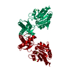 6cfrS S: Starting model for refinement |
|---|---|
| Similar structure data |
-Links
 PDBj
PDBj- Assembly
| Deposited unit | 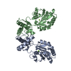
| ||||||||
|---|---|---|---|---|---|---|---|---|---|
| 1 | 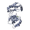
| ||||||||
| 2 | 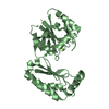
| ||||||||
| Unit cell |
|
-Components
| #1: Protein | Mass: 28921.934 Da / Num. of mol.: 2 Source method: isolated from a genetically manipulated source Source: (gene. exp.) Strain: isolate 3D7 / Gene: PF3D7_1017400 / Plasmid: pET28a / Production host:  #2: Chemical |   Mass: 24.305 Da / Num. of mol.: 2 / Source method: obtained synthetically / Formula: Mg Mass: 24.305 Da / Num. of mol.: 2 / Source method: obtained synthetically / Formula: MgHas ligand of interest | N | |
|---|
-Experimental details
-Experiment
| Experiment | Method: X-RAY DIFFRACTION / Number of used crystals: 1 |
|---|
- Sample preparation
| Crystal | Density Matthews: 3.56 Å3/Da / Density % sol: 65.45 % |
|---|---|
| Crystal grow | Temperature: 277 K / Method: vapor diffusion, hanging drop / pH: 6.5 Details: 2ul drops containing 1:1 mixture of protein (6mg/mL) and crystallization buffer (0.1M bis -tris-propane pH 6.5, 0.1 M calcium acetate, 16% (w/v) PEG 8000, and 2% (v/v) benzamidine hydrochloride) |
-Data collection
| Diffraction | Mean temperature: 100 K / Serial crystal experiment: N |
|---|---|
| Diffraction source | Source: SYNCHROTRON / Site: APS / Beamline: 19-ID / Wavelength: 0.98 Å |
| Detector | Type: DECTRIS PILATUS3 X 6M / Detector: PIXEL / Date: Jul 30, 2020 |
| Radiation | Protocol: SINGLE WAVELENGTH / Monochromatic (M) / Laue (L): M / Scattering type: x-ray |
| Radiation wavelength | Wavelength: 0.98 Å / Relative weight: 1 |
| Reflection | Resolution: 3.5→40.3164 Å / Num. obs: 11030 / % possible obs: 99.9 % / Redundancy: 32.1 % / Rsym value: 0.334 / Net I/σ(I): 18.5 |
| Reflection shell | Resolution: 3.5→3.56 Å / Redundancy: 30.1 % / Mean I/σ(I) obs: 2.7 / Num. unique obs: 543 / Rsym value: 1.43 / % possible all: 100 |
- Processing
| Software |
| |||||||||||||||||||||||||||||||||||||||||||||||||||||||||||||||
|---|---|---|---|---|---|---|---|---|---|---|---|---|---|---|---|---|---|---|---|---|---|---|---|---|---|---|---|---|---|---|---|---|---|---|---|---|---|---|---|---|---|---|---|---|---|---|---|---|---|---|---|---|---|---|---|---|---|---|---|---|---|---|---|---|
| Refinement | Method to determine structure: MOLECULAR REPLACEMENT Starting model: 6CFR Resolution: 3.51→40.31 Å / SU ML: 0.5 / Cross valid method: THROUGHOUT / σ(F): 1.38 / Phase error: 31.24 / Stereochemistry target values: ML
| |||||||||||||||||||||||||||||||||||||||||||||||||||||||||||||||
| Solvent computation | Shrinkage radii: 0.9 Å / VDW probe radii: 1.11 Å / Solvent model: FLAT BULK SOLVENT MODEL | |||||||||||||||||||||||||||||||||||||||||||||||||||||||||||||||
| Displacement parameters | Biso max: 164.07 Å2 / Biso mean: 86.4114 Å2 / Biso min: 44.48 Å2 | |||||||||||||||||||||||||||||||||||||||||||||||||||||||||||||||
| Refinement step | Cycle: final / Resolution: 3.51→40.31 Å
| |||||||||||||||||||||||||||||||||||||||||||||||||||||||||||||||
| LS refinement shell | Refine-ID: X-RAY DIFFRACTION / Rfactor Rfree error: 0 / Total num. of bins used: 8
|
