 Movie
Movie Controller

[English] 日本語
 Yorodumi
- PDB-7myd: Leishmania major dihydroorotate dehydrogenase in complex with 5-a... -
+ Open data

- Basic information
| Entry | Database: PDB / ID: 7myd | ||||||
|---|---|---|---|---|---|---|---|
| Title | Leishmania major dihydroorotate dehydrogenase in complex with 5-amino-2-(1H-pyrrol-1-yl)benzonitrile | ||||||
 Components Components | Dihydroorotate dehydrogenase (fumarate) | ||||||
 Keywords Keywords | OXIDOREDUCTASE / Leishmania major / neglected tropical disease / dihydroorotate dehydrogenase | ||||||
| Function / homology |  Function and homology information Function and homology informationdihydroorotate dehydrogenase (fumarate) / dihydroorotate dehydrogenase (fumarate) activity / fumarate metabolic process / dihydroorotate dehydrogenase activity / ciliary plasm / 'de novo' UMP biosynthetic process / 'de novo' pyrimidine nucleobase biosynthetic process / nucleotide binding / nucleoplasm / cytoplasm Similarity search - Function | ||||||
| Biological species |  Leishmania major (eukaryote) Leishmania major (eukaryote) | ||||||
| Method |  X-RAY DIFFRACTION / SYNCHROTRON / MOLECULAR REPLACEMENT / Resolution: 2.15 Å X-RAY DIFFRACTION / SYNCHROTRON / MOLECULAR REPLACEMENT / Resolution: 2.15 Å | ||||||
 Authors Authors | Pinheiro, M.P. / Cardoso, I.A. / Hunter, W.N. / Nonato, M.C. | ||||||
| Funding support |  Brazil, 1items Brazil, 1items
| ||||||
 Citation Citation | Journal: To Be Published Title: Leishmania major dihydroorotate dehydrogenase in complex with 5-amino-2-(1H-pyrrol-1-yl)benzonitrile Authors: Pinheiro, M.P. / Cardoso, I.A. / Hunter, W.N. / Nonato, M.C. | ||||||
| History |
|
- Structure visualization
| Structure viewer | Molecule: MolmilJmol/JSmol |
|---|
- Downloads & links
-Download
| PDBx/mmCIF format | 7myd.cif.gz | 254.8 KB | Display | PDBx/mmCIF format |
|---|---|---|---|---|
| PDB format | pdb7myd.ent.gz | Display | PDB format | |
| PDBx/mmJSON format | 7myd.json.gz | Tree view | PDBx/mmJSON format | |
| Others |  Other downloads Other downloads |
-Validation report
| Arichive directory | https://data.pdbj.org/pub/pdb/validation_reports/my/7mydftp://data.pdbj.org/pub/pdb/validation_reports/my/7myd | HTTPS FTP |
|---|
-Related structure data
| Related structure data | 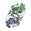 3gyeS S: Starting model for refinement |
|---|---|
| Similar structure data |
-Links
 PDBj
PDBj- Assembly
| Deposited unit | 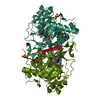
| ||||||||
|---|---|---|---|---|---|---|---|---|---|
| 1 |
| ||||||||
| Unit cell |
|
-Components
-Protein , 1 types, 2 molecules AAABBB
| #1: Protein | Mass: 38245.641 Da / Num. of mol.: 2 Source method: isolated from a genetically manipulated source Source: (gene. exp.) Leishmania major (eukaryote) / Gene: DHODH, LMJF_16_0530 / Plasmid: pET28A / Production host:  References: UniProt: Q4QEW7, dihydroorotate dehydrogenase (fumarate) |
|---|
-Non-polymers , 5 types, 343 molecules 


| #2: Chemical |  Mass: 456.344 Da / Num. of mol.: 2 / Source method: obtained synthetically / Formula: C17H21N4O9P Mass: 456.344 Da / Num. of mol.: 2 / Source method: obtained synthetically / Formula: C17H21N4O9P#3: Chemical |  Mass: 96.063 Da / Num. of mol.: 3 / Source method: obtained synthetically / Formula: SO4 Mass: 96.063 Da / Num. of mol.: 3 / Source method: obtained synthetically / Formula: SO4#4: Chemical |  Mass: 92.094 Da / Num. of mol.: 3 / Source method: obtained synthetically / Formula: C3H8O3 Mass: 92.094 Da / Num. of mol.: 3 / Source method: obtained synthetically / Formula: C3H8O3#5: Chemical | ChemComp-SNQ / |  Mass: 183.209 Da / Num. of mol.: 1 / Source method: obtained synthetically / Formula: C11H9N3 / Feature type: SUBJECT OF INVESTIGATION Mass: 183.209 Da / Num. of mol.: 1 / Source method: obtained synthetically / Formula: C11H9N3 / Feature type: SUBJECT OF INVESTIGATION#6: Water | ChemComp-HOH / | Mass: 18.015 Da / Num. of mol.: 334 / Source method: isolated from a natural source / Formula: H2O |
|---|
-Details
| Has ligand of interest | Y |
|---|
-Experimental details
-Experiment
| Experiment | Method: X-RAY DIFFRACTION / Number of used crystals: 1 |
|---|
- Sample preparation
| Crystal | Density Matthews: 2.67 Å3/Da / Density % sol: 53.85 % |
|---|---|
| Crystal grow | Temperature: 295 K / Method: vapor diffusion, sitting drop Details: 100 mM sodium citrate tribasic dihydrate, pH 5.6, 1.3 M lithium sulfate, 0.30 M ammonium sulfate |
-Data collection
| Diffraction | Mean temperature: 100 K / Serial crystal experiment: N |
|---|---|
| Diffraction source | Source: SYNCHROTRON / Site: LNLS / Beamline: D03B-MX1 / Wavelength: 1.437 Å |
| Detector | Type: MAR CCD 165 mm / Detector: CCD / Date: Apr 15, 2011 |
| Radiation | Protocol: SINGLE WAVELENGTH / Monochromatic (M) / Laue (L): M / Scattering type: x-ray |
| Radiation wavelength | Wavelength: 1.437 Å / Relative weight: 1 |
| Reflection | Resolution: 2.15→35.77 Å / Num. obs: 43015 / % possible obs: 97.8 % / Redundancy: 7 % / Rmerge(I) obs: 0.137 / Rsym value: 0.137 / Net I/σ(I): 11.9 |
| Reflection shell | Resolution: 2.15→2.27 Å / Redundancy: 6.7 % / Rmerge(I) obs: 0.803 / Mean I/σ(I) obs: 2.6 / Num. unique obs: 6253 / Rsym value: 0.803 / % possible all: 98.1 |
- Processing
| Software |
| |||||||||||||||||||||||||||||||||||||||||||||||||||||||||||||||||||||||||||||||||||||||||||||||||||||||||||||||||||||||||||||||||||||||||||||||||||||||||||
|---|---|---|---|---|---|---|---|---|---|---|---|---|---|---|---|---|---|---|---|---|---|---|---|---|---|---|---|---|---|---|---|---|---|---|---|---|---|---|---|---|---|---|---|---|---|---|---|---|---|---|---|---|---|---|---|---|---|---|---|---|---|---|---|---|---|---|---|---|---|---|---|---|---|---|---|---|---|---|---|---|---|---|---|---|---|---|---|---|---|---|---|---|---|---|---|---|---|---|---|---|---|---|---|---|---|---|---|---|---|---|---|---|---|---|---|---|---|---|---|---|---|---|---|---|---|---|---|---|---|---|---|---|---|---|---|---|---|---|---|---|---|---|---|---|---|---|---|---|---|---|---|---|---|---|---|---|
| Refinement | Method to determine structure: MOLECULAR REPLACEMENT Starting model: PDB entry 3GYE Resolution: 2.15→35.77 Å / Cor.coef. Fo:Fc: 0.954 / Cor.coef. Fo:Fc free: 0.927 / SU B: 4.971 / SU ML: 0.124 / Cross valid method: FREE R-VALUE / ESU R: 0.197 / ESU R Free: 0.171 Details: Hydrogens have been added in their riding positions
| |||||||||||||||||||||||||||||||||||||||||||||||||||||||||||||||||||||||||||||||||||||||||||||||||||||||||||||||||||||||||||||||||||||||||||||||||||||||||||
| Solvent computation | Ion probe radii: 0.8 Å / Shrinkage radii: 0.8 Å / VDW probe radii: 1.2 Å / Solvent model: MASK BULK SOLVENT | |||||||||||||||||||||||||||||||||||||||||||||||||||||||||||||||||||||||||||||||||||||||||||||||||||||||||||||||||||||||||||||||||||||||||||||||||||||||||||
| Displacement parameters | Biso mean: 26.369 Å2
| |||||||||||||||||||||||||||||||||||||||||||||||||||||||||||||||||||||||||||||||||||||||||||||||||||||||||||||||||||||||||||||||||||||||||||||||||||||||||||
| Refinement step | Cycle: LAST / Resolution: 2.15→35.77 Å
| |||||||||||||||||||||||||||||||||||||||||||||||||||||||||||||||||||||||||||||||||||||||||||||||||||||||||||||||||||||||||||||||||||||||||||||||||||||||||||
| Refine LS restraints |
| |||||||||||||||||||||||||||||||||||||||||||||||||||||||||||||||||||||||||||||||||||||||||||||||||||||||||||||||||||||||||||||||||||||||||||||||||||||||||||
| LS refinement shell |
|
