 Movie
Movie Controller


+ Open data

- Basic information
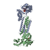
| Entry | Database: PDB / ID: 7msk | ||||||
|---|---|---|---|---|---|---|---|
| Title | ThuS glycosin S-glycosyltransferase | ||||||
 Components Components | Glyco_trans_2-like domain-containing protein | ||||||
 Keywords Keywords | BIOSYNTHETIC PROTEIN / glycosin / RiPP / glycosyltransferase | ||||||
| Function / homology | : / Peptide S-glycosyltransferase, SunS family / Glycosyltransferase 2-like / Glycosyl transferase family 2 / Nucleotide-diphospho-sugar transferases / Chem-U2F / Glyco_trans_2-like domain-containing protein Function and homology information Function and homology information | ||||||
| Biological species |  | ||||||
| Method |  X-RAY DIFFRACTION / SYNCHROTRON / MOLECULAR REPLACEMENT / Resolution: 2.06 Å X-RAY DIFFRACTION / SYNCHROTRON / MOLECULAR REPLACEMENT / Resolution: 2.06 Å | ||||||
 Authors Authors | Garg, N. / Nair, S.K. | ||||||
| Funding support |  United States, 1items United States, 1items
| ||||||
 Citation Citation | Journal: Cell Chem Biol / Year: 2021 Title: Structural and mechanistic investigations of protein S-glycosyltransferases. Authors: Fujinami, D. / Garcia de Gonzalo, C.V. / Biswas, S. / Hao, Y. / Wang, H. / Garg, N. / Lukk, T. / Nair, S.K. / van der Donk, W.A. | ||||||
| History |
|
- Structure visualization
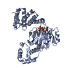
| Structure viewer | Molecule: MolmilJmol/JSmol |
|---|
- Downloads & links
-Download
| PDBx/mmCIF format | 7msk.cif.gz | 195 KB | Display | PDBx/mmCIF format |
|---|---|---|---|---|
| PDB format | pdb7msk.ent.gz | 152.5 KB | Display | PDB format |
| PDBx/mmJSON format | 7msk.json.gz | Tree view | PDBx/mmJSON format | |
| Others |  Other downloads Other downloads |
-Validation report
| Summary document | 7msk_validation.pdf.gz | 1.3 MB | Display | wwPDB validaton report |
|---|---|---|---|---|
| Full document | 7msk_full_validation.pdf.gz | 1.3 MB | Display | |
| Data in XML | 7msk_validation.xml.gz | 34.4 KB | Display | |
| Data in CIF | 7msk_validation.cif.gz | 48.9 KB | Display | |
| Arichive directory | https://data.pdbj.org/pub/pdb/validation_reports/ms/7mskftp://data.pdbj.org/pub/pdb/validation_reports/ms/7msk | HTTPS FTP |
-Related structure data
-Links
 PDBj
PDBj

- Assembly
| Deposited unit | 
| ||||||||
|---|---|---|---|---|---|---|---|---|---|
| 1 |
| ||||||||
| Unit cell |
|
-Components
| #1: Protein | Mass: 50454.461 Da / Num. of mol.: 2 Source method: isolated from a genetically manipulated source Source: (gene. exp.) Gene: bthur0009_56280 / Production host: #2: Chemical |   Mass: 24.305 Da / Num. of mol.: 2 / Source method: obtained synthetically / Formula: Mg / Feature type: SUBJECT OF INVESTIGATION Mass: 24.305 Da / Num. of mol.: 2 / Source method: obtained synthetically / Formula: Mg / Feature type: SUBJECT OF INVESTIGATION#3: Chemical | 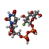  Mass: 568.293 Da / Num. of mol.: 2 / Source method: obtained synthetically / Formula: C15H23FN2O16P2 / Feature type: SUBJECT OF INVESTIGATION Mass: 568.293 Da / Num. of mol.: 2 / Source method: obtained synthetically / Formula: C15H23FN2O16P2 / Feature type: SUBJECT OF INVESTIGATION#4: Water | ChemComp-HOH / |  Mass: 18.015 Da / Num. of mol.: 337 / Source method: isolated from a natural source / Formula: H2O Mass: 18.015 Da / Num. of mol.: 337 / Source method: isolated from a natural source / Formula: H2OHas ligand of interest | Y | |
|---|
-Experimental details
-Experiment
| Experiment | Method: X-RAY DIFFRACTION / Number of used crystals: 1 |
|---|
- Sample preparation
| Crystal | Density Matthews: 2.51 Å3/Da / Density % sol: 51.04 % |
|---|---|
| Crystal grow | Temperature: 283 K / Method: vapor diffusion, hanging drop Details: 15% PEG 3350 200 mM ammonium nitrate 100 mM BisTris HCl pH=6.5 |
-Data collection
| Diffraction | Mean temperature: 100 K / Serial crystal experiment: N |
|---|---|
| Diffraction source | Source: SYNCHROTRON / Site: APS / Beamline: 21-ID-G / Wavelength: 0.9786 Å |
| Detector | Type: MAR scanner 300 mm plate / Detector: IMAGE PLATE / Date: Dec 19, 2019 |
| Radiation | Protocol: SINGLE WAVELENGTH / Monochromatic (M) / Laue (L): M / Scattering type: x-ray |
| Radiation wavelength | Wavelength: 0.9786 Å / Relative weight: 1 |
| Reflection | Resolution: 2.06→90.3 Å / Num. obs: 61544 / % possible obs: 99.7 % / Redundancy: 6.3 % / CC1/2: 0.999 / Net I/σ(I): 16.9 |
| Reflection shell | Resolution: 2.06→2.09 Å / Num. unique obs: 3042 / CC1/2: 0.831 |
- Processing
| Software |
| ||||||||||||||||||||||||||||||||||||||||||||||||||||||||||||
|---|---|---|---|---|---|---|---|---|---|---|---|---|---|---|---|---|---|---|---|---|---|---|---|---|---|---|---|---|---|---|---|---|---|---|---|---|---|---|---|---|---|---|---|---|---|---|---|---|---|---|---|---|---|---|---|---|---|---|---|---|---|
| Refinement | Method to determine structure: MOLECULAR REPLACEMENT / Resolution: 2.06→25 Å / Cor.coef. Fo:Fc: 0.952 / Cor.coef. Fo:Fc free: 0.933 / SU B: 5.602 / SU ML: 0.146 / Cross valid method: THROUGHOUT / σ(F): 0 / ESU R: 0.213 / ESU R Free: 0.178 / Stereochemistry target values: MAXIMUM LIKELIHOOD Details: HYDROGENS HAVE BEEN USED IF PRESENT IN THE INPUT U VALUES : REFINED INDIVIDUALLY
| ||||||||||||||||||||||||||||||||||||||||||||||||||||||||||||
| Solvent computation | Ion probe radii: 0.8 Å / Shrinkage radii: 0.8 Å / VDW probe radii: 1.2 Å / Solvent model: MASK | ||||||||||||||||||||||||||||||||||||||||||||||||||||||||||||
| Displacement parameters | Biso max: 113.68 Å2 / Biso mean: 41.5 Å2 / Biso min: 19.11 Å2
| ||||||||||||||||||||||||||||||||||||||||||||||||||||||||||||
| Refinement step | Cycle: final / Resolution: 2.06→25 Å
| ||||||||||||||||||||||||||||||||||||||||||||||||||||||||||||
| Refine LS restraints |
| ||||||||||||||||||||||||||||||||||||||||||||||||||||||||||||
| LS refinement shell | Resolution: 2.06→2.113 Å / Rfactor Rfree error: 0 / Total num. of bins used: 20
|
