 Movie
Movie Controller

[English] 日本語
 Yorodumi
- PDB-7mod: Crystal Structure of Arabidopsis thaliana Plant and Fungi Atypica... -
+ Open data

- Basic information
| Entry | Database: PDB / ID: 7mod | ||||||
|---|---|---|---|---|---|---|---|
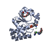
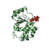
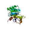
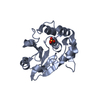
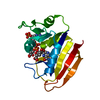
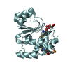
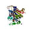
| Title | Crystal Structure of Arabidopsis thaliana Plant and Fungi Atypical Dual Specificity Phosphatase 1(AtPFA-DSP1 ) Cys150Ser in Complex with Phosphate in Conformation A (Pi(A)) | ||||||
 Components Components | Tyrosine-protein phosphatase DSP1 | ||||||
 Keywords Keywords | HYDROLASE / inositol / inositol pyrophosphate / TRANSFERASE / cell-signaling / phosphatase / substrate recognition / reaction mechanism / intermediate / phosphate / metaphosphate / molecular dynamic simulation / self-activation / catalytic water | ||||||
| Function / homology |  Function and homology information Function and homology informationinositol-1,5-bisdiphosphate-2,3,4,6-tetrakisphosphate 5-diphosphatase activity / inositol-5-diphosphate-1,3,4,6-tetrakisphosphate diphosphatase activity / inositol-3,5-bisdiphosphate-2,3,4,6-tetrakisphosphate 5-diphosphatase activity / inositol-5-diphosphate-1,2,3,4,6-pentakisphosphate diphosphatase activity / diphosphoinositol-polyphosphate diphosphatase / phosphatase activity / cytoplasm Similarity search - Function | ||||||
| Biological species |  | ||||||
| Method |  X-RAY DIFFRACTION / SYNCHROTRON / MOLECULAR REPLACEMENT / Resolution: 1.65 Å X-RAY DIFFRACTION / SYNCHROTRON / MOLECULAR REPLACEMENT / Resolution: 1.65 Å | ||||||
 Authors Authors | Wang, H. / Shears, S.B. | ||||||
| Funding support |  United States, 1items United States, 1items
| ||||||
 Citation Citation | Journal: Nat Commun / Year: 2022 Title: A structural expose of noncanonical molecular reactivity within the protein tyrosine phosphatase WPD loop. Authors: Wang, H. / Perera, L. / Jork, N. / Zong, G. / Riley, A.M. / Potter, B.V.L. / Jessen, H.J. / Shears, S.B. | ||||||
| History |
|
- Structure visualization
| Structure viewer | Molecule: MolmilJmol/JSmol |
|---|
- Downloads & links
-Download
| PDBx/mmCIF format | 7mod.cif.gz | 170.4 KB | Display | PDBx/mmCIF format |
|---|---|---|---|---|
| PDB format | pdb7mod.ent.gz | 134.3 KB | Display | PDB format |
| PDBx/mmJSON format | 7mod.json.gz | Tree view | PDBx/mmJSON format | |
| Others |  Other downloads Other downloads |
-Validation report
| Arichive directory | https://data.pdbj.org/pub/pdb/validation_reports/mo/7modftp://data.pdbj.org/pub/pdb/validation_reports/mo/7mod | HTTPS FTP |
|---|
-Related structure data
| Related structure data | 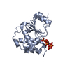 7moeC  7mofC  7mogC  7mohC  7moiC 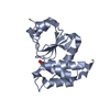 7mojC  7mokC  7molC  7momC 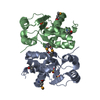 1xriS S: Starting model for refinement C: citing same article ( |
|---|---|
| Similar structure data |


-Links
 PDBj
PDBj


- Assembly
| Deposited unit | 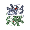
| |||||||||
|---|---|---|---|---|---|---|---|---|---|---|
| 1 | 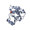
| |||||||||
| 2 | 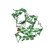
| |||||||||
| Unit cell |
| |||||||||
| Components on special symmetry positions |
|
-Components
| #1: Protein | Mass: 19642.770 Da / Num. of mol.: 2 / Mutation: C150S Source method: isolated from a genetically manipulated source Source: (gene. exp.)  #2: Chemical |   Mass: 94.971 Da / Num. of mol.: 2 / Source method: obtained synthetically / Formula: PO4 / Feature type: SUBJECT OF INVESTIGATION Mass: 94.971 Da / Num. of mol.: 2 / Source method: obtained synthetically / Formula: PO4 / Feature type: SUBJECT OF INVESTIGATION#3: Water | ChemComp-HOH / |  Mass: 18.015 Da / Num. of mol.: 416 / Source method: isolated from a natural source / Formula: H2O Mass: 18.015 Da / Num. of mol.: 416 / Source method: isolated from a natural source / Formula: H2OHas ligand of interest | Y | |
|---|
-Experimental details
-Experiment
| Experiment | Method: X-RAY DIFFRACTION / Number of used crystals: 1 |
|---|
- Sample preparation
| Crystal | Density Matthews: 4.18 Å3/Da / Density % sol: 70.56 % |
|---|---|
| Crystal grow | Temperature: 298 K / Method: vapor diffusion, hanging drop Details: 0.4 M NaCl, 100 mM HEPES pH7.2, 50 mM beta-mercaptoethanol at 298K (3 ul of 5.5 mg/ml protein plus 1 ul of well buffer in the crystallization drop). |
-Data collection
| Diffraction | Mean temperature: 100 K / Serial crystal experiment: N |
|---|---|
| Diffraction source | Source: SYNCHROTRON / Site: APS / Beamline: 22-BM / Wavelength: 1 Å |
| Detector | Type: MARMOSAIC 300 mm CCD / Detector: CCD / Date: Oct 6, 2017 |
| Radiation | Protocol: SINGLE WAVELENGTH / Monochromatic (M) / Laue (L): M / Scattering type: x-ray |
| Radiation wavelength | Wavelength: 1 Å / Relative weight: 1 |
| Reflection | Resolution: 1.65→50 Å / Num. obs: 74329 / % possible obs: 99.6 % / Redundancy: 7.8 % / Rrim(I) all: 0.076 / Net I/σ(I): 27.5 |
| Reflection shell | Resolution: 1.65→1.69 Å / Mean I/σ(I) obs: 2.5 / Num. unique obs: 5335 / Rrim(I) all: 0.849 |
- Processing
| Software |
| |||||||||||||||||||||||||||||||||||||||||||||||||||||||||||||||||
|---|---|---|---|---|---|---|---|---|---|---|---|---|---|---|---|---|---|---|---|---|---|---|---|---|---|---|---|---|---|---|---|---|---|---|---|---|---|---|---|---|---|---|---|---|---|---|---|---|---|---|---|---|---|---|---|---|---|---|---|---|---|---|---|---|---|---|
| Refinement | Method to determine structure: MOLECULAR REPLACEMENT Starting model: 1XRI Resolution: 1.65→44.36 Å / Cor.coef. Fo:Fc: 0.978 / Cor.coef. Fo:Fc free: 0.968 / SU B: 2.02 / SU ML: 0.03 / Cross valid method: THROUGHOUT / σ(F): 0 / ESU R: 0.052 / ESU R Free: 0.05 / Stereochemistry target values: MAXIMUM LIKELIHOOD Details: HYDROGENS HAVE BEEN ADDED IN THE RIDING POSITIONS U VALUES : REFINED INDIVIDUALLY
| |||||||||||||||||||||||||||||||||||||||||||||||||||||||||||||||||
| Solvent computation | Ion probe radii: 0.8 Å / Shrinkage radii: 0.8 Å / VDW probe radii: 1.2 Å / Solvent model: MASK | |||||||||||||||||||||||||||||||||||||||||||||||||||||||||||||||||
| Displacement parameters | Biso max: 343.37 Å2 / Biso mean: 20.841 Å2 / Biso min: 5.27 Å2
| |||||||||||||||||||||||||||||||||||||||||||||||||||||||||||||||||
| Refinement step | Cycle: final / Resolution: 1.65→44.36 Å
| |||||||||||||||||||||||||||||||||||||||||||||||||||||||||||||||||
| Refine LS restraints |
| |||||||||||||||||||||||||||||||||||||||||||||||||||||||||||||||||
| LS refinement shell | Resolution: 1.651→1.694 Å / Rfactor Rfree error: 0 / Total num. of bins used: 20
|
