| 登録情報 | データベース: PDB / ID: 7mku
|
|---|
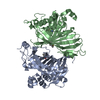
| タイトル | Crystal Structure of ENOYL COA-HYDRATASE2 from Arabidopsis thaliana |
|---|
 要素 要素 | Enoyl-CoA hydratase 2, peroxisomal |
|---|
 キーワード キーワード | LYASE / hydratase / hot-dog fold / PLANT PROTEIN |
|---|
| 機能・相同性 |  機能・相同性情報 機能・相同性情報
fatty acid beta-oxidation, unsaturated, even number / enoyl-CoA hydratase 2 / peroxisome / oxidoreductase activity / lyase activity / mitochondrion類似検索 - 分子機能 : / MFE-2 hydratase 2 N-terminal domain / MaoC-like dehydratase domain / MaoC like domain / HotDog domain superfamily類似検索 - ドメイン・相同性 |
|---|
| 生物種 |   Arabidopsis thaliana (シロイヌナズナ) Arabidopsis thaliana (シロイヌナズナ) |
|---|
| 手法 |  X線回折 / シンクロトロン / 単波長異常分散 / 解像度: 2.648 Å X線回折 / シンクロトロン / 単波長異常分散 / 解像度: 2.648 Å |
|---|
 データ登録者 データ登録者 | Power, S.K. / Korasick, D.A. / Jez, J.M. / Strader, L.C. |
|---|
| 資金援助 |  米国, 5件 米国, 5件 | 組織 | 認可番号 | 国 |
|---|
| National Institutes of Health/National Institute of General Medical Sciences (NIH/NIGMS) | R35 GM136338 | 米国 | | National Science Foundation (NSF, United States) | IOS-1453750 | 米国 | | National Science Foundation (NSF, United States) | MCB-1614539 | 米国 | | United States Department of Agriculture (USDA) | MOW-2014-01877 | 米国 | | National Science Foundation (NSF, United States) | 2011101911 | 米国 |
|
|---|
 引用 引用 | ジャーナル: To Be Published
タイトル: Crystal Structure of ENOYL COA-HYDRATASE2 from Arabidopsis thaliana
著者: Korasick, D.A. / Powers, S.K. / Jez, J.M. / Strader, L.C. |
|---|
| 履歴 | | 登録 | 2021年4月26日 | 登録サイト: RCSB / 処理サイト: RCSB |
|---|
| 改定 1.0 | 2022年5月11日 | Provider: repository / タイプ: Initial release |
|---|
| 改定 1.1 | 2024年11月20日 | Group: Data collection / Refinement description / Structure summary
カテゴリ: chem_comp_atom / chem_comp_bond ...chem_comp_atom / chem_comp_bond / pdbx_entry_details / pdbx_modification_feature / struct_ncs_dom_lim
Item: _pdbx_entry_details.has_protein_modification / _struct_ncs_dom_lim.beg_auth_comp_id ..._pdbx_entry_details.has_protein_modification / _struct_ncs_dom_lim.beg_auth_comp_id / _struct_ncs_dom_lim.beg_label_asym_id / _struct_ncs_dom_lim.beg_label_comp_id / _struct_ncs_dom_lim.beg_label_seq_id / _struct_ncs_dom_lim.end_auth_comp_id / _struct_ncs_dom_lim.end_label_asym_id / _struct_ncs_dom_lim.end_label_comp_id / _struct_ncs_dom_lim.end_label_seq_id |
|---|
|
|---|
 ムービー
ムービー コントローラー


 データを開く
 基本情報
基本情報 構造の表示
構造の表示 ダウンロードとリンク
 その他のダウンロード
 PDBj
PDBj
 集合体
集合体